
Page 609 - Atlas of Small Animal CT and MRI
P. 609
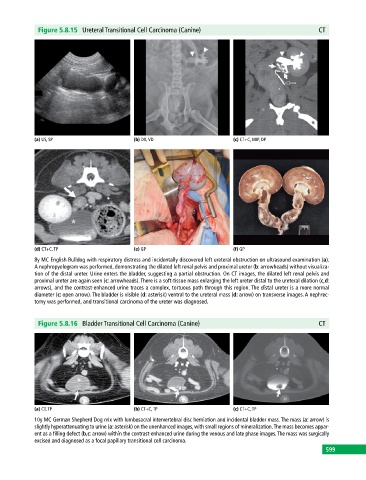
Figure 5.8.15  Ureteral Transitional Cell Carcinoma (Canine)                                CT


            (a) US, SP                       (b) DX, VD                       (c) CT+C, MIP, DP


            (d) CT+C, TP                     (e) GP                           (f) GP
            8y MC English Bulldog with respiratory distress and incidentally discovered left ureteral obstruction on ultrasound examination (a).
            A nephropyelogram was performed, demonstrating the dilated left renal pelvis and proximal ureter (b: arrowheads) without visualiza-
            tion of the distal ureter. Urine enters the bladder, suggesting a partial obstruction. On CT images, the dilated left renal pelvis and
            proximal ureter are again seen (c: arrowheads). There is a soft‐tissue mass enlarging the left ureter distal to the ureteral dilation (c,d:
            arrows), and the contrast‐enhanced urine traces a complex, tortuous path through this region. The distal ureter is a more normal
            diameter (c: open arrow). The bladder is visible (d: asterisk) ventral to the ureteral mass (d: arrow) on transverse images. A nephrec-
            tomy was performed, and transitional carcinoma of the ureter was diagnosed.


              Figure 5.8.16  Bladder Transitional Cell Carcinoma (Canine)                                 CT


            (a) CT, TP                       (b) CT+C, TP                     (c) CT+C, TP
            10y MC German Shepherd Dog mix with lumbosacral intervertebral disc herniation and incidental bladder mass. The mass (a: arrow) is
            slightly hyperattenuating to urine (a: asterisk) on the unenhanced images, with small regions of mineralization. The mass becomes appar-
            ent as a filling defect (b,c: arrow) within the contrast‐enhanced urine during the venous and late phase images. The mass was surgically
            excised and diagnosed as a focal papillary transitional cell carcinoma.
                                                                                                             599