
Page 146 - Atlas of Small Animal CT and MRI
P. 146
136 Atlas of Small Animal CT and MRI
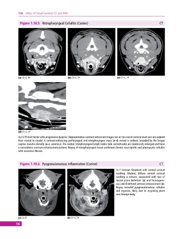
Figure 1.10.5 Retropharyngeal Cellulitis (Canine) CT
(a) CT+C, TP (b) CT+C, TP (c) CT+C, TP
(d) CT+C, SP
4y FS Pit Bull Terrier with progressive dyspnea. Representative contrast‐enhanced images are at the cranial cervical level and are ordered
from cranial to caudal. A contrast‐enhancing perilaryngeal and retropharyngeal mass (a–d: arrow) is evident, bounded by the longus
capitus muscles dorsally (a–c: asterisks). The medial retropharyngeal lymph nodes (a,b: arrowheads) are moderately enlarged and have
a nonuniform contrast‐enhancement pattern. Biopsy of retropharyngeal tissue confirmed chronic neutrophilic and plasmacytic cellulitis
with extensive fibrosis.
Figure 1.10.6 Pyogranulomatous Inflammation (Canine) CT
2y F German Shepherd with ventral cervical
swelling. Marked, diffuse ventral cervical
swelling is present, associated with loss of
fascial plane definition (a) and heterogene-
ous and ill‐defined contrast enhancement (b).
Biopsy revealed pyogranulomatous cellulitis
and myositis, likely due to migrating plant
awn foreign body.
(a) CT, TP (b) CT+C, TP
136