
Page 147 - Atlas of Small Animal CT and MRI
P. 147
Larynx, Pharynx, and Neck 137
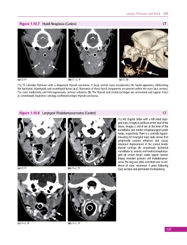
Figure 1.10.7 Hyoid Neoplasia (Canine) CT
(a) CT, TP (b) CT+C, TP (c) CT, 3D
11y FS Labrador Retriever with a diagnosed thyroid carcinoma. A large ventral mass incorporates the hyoid apparatus, obliterating
the basihyoid, thyrohyoid, and ceratohyoid bones (a,c). Remnants of these hyoid components are present within the mass (a,c: arrows).
The mass moderately and heterogeneously contrast enhances (b). The thyroid and cricoid cartilages are uninvolved and appear intact
(c: arrowhead). Aspiration cytology confirmed ectopic thyroid carcinoma.
Figure 1.10.8 Laryngeal Rhabdomyosarcoma (Canine) CT
11y MC English Setter with a left‐sided laryn-
geal mass. Images a and b are at the level of the
larynx. Images c and d are at the level of the
mandibular and medial retropharyngeal lymph
nodes, respectively. There is a centrally hypoat-
tenuating left laryngeal mass (a,b: arrow) that
peripherally contrast enhances and causes
rotational displacement of the cranial border
thyroid cartilage (b: arrowhead). Ipsilateral
mandibular (c: arrows) and medial retropharyn-
geal (d: arrow) lymph nodes appear normal.
Biopsy revealed granular cell rhabdomyosar-
coma. The dog was alive, and there was no evi-
dence of mass recurrence 4 years following
(a) CT, TP (b) CT+C, TP mass excision and permanent tracheostomy.
(c) CT+C, TP (d) CT+C, TP
137