
Page 144 - Atlas of Small Animal CT and MRI
P. 144
134 Atlas of Small Animal CT and MRI
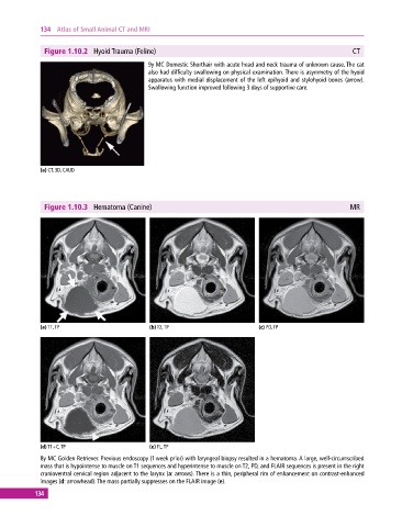
Figure 1.10.2 Hyoid Trauma (Feline) CT
9y MC Domestic Shorthair with acute head and neck trauma of unknown cause. The cat
also had difficulty swallowing on physical examination. There is asymmetry of the hyoid
apparatus with medial displacement of the left epihyoid and stylohyoid bones (arrow).
Swallowing function improved following 3 days of supportive care.
(a) CT, 3D, CAUD
Figure 1.10.3 Hematoma (Canine) MR
(a) T1, TP (b) T2, TP (c) PD, TP
(d) T1+C, TP (e) FL, TP
8y MC Golden Retriever. Previous endoscopy (1 week prior) with laryngeal biopsy resulted in a hematoma. A large, well‐circumscribed
mass that is hypointense to muscle on T1 sequences and hyperintense to muscle on T2, PD, and FLAIR sequences is present in the right
cranioventral cervical region adjacent to the larynx (a: arrows). There is a thin, peripheral rim of enhancement on contrast‐enhanced
images (d: arrowhead). The mass partially suppresses on the FLAIR image (e).
134