
Page 148 - Atlas of Small Animal CT and MRI
P. 148
138 Atlas of Small Animal CT and MRI
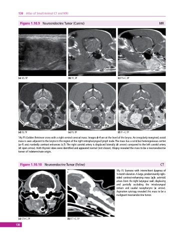
Figure 1.10.9 Neuroendocrine Tumor (Canine) MR
(a) US, SP (b) T1, SP (c) T1+C, SP
(d) T2, TP (e) T1, TP (f) T1+C, TP
14y FS Golden Retriever cross with a right ventral cervical mass. Images d–f are at the level of the larynx. An irregularly margined, ovoid
mass is seen adjacent to the larynx in the region of the right retropharyngeal lymph node. The mass has a solid but heterogeneous center
(a–f) and markedly contrast enhances (c,f). The right carotid artery is displaced laterally (d: arrow) compared to the left carotid artery
(d: open arrow). Both thyroid lobes were identified and appeared normal (not shown). Biopsy revealed the mass to be a neuroendocrine
tumor of indeterminate origin.
Figure 1.10.10 Neuroendocrine Tumor (Feline) CT
10y FS Siamese with intermittent dyspnea of
1‐month duration. A large, predominantly right‐
sided contrast‐enhancing mass (a,b: asterisk)
arises from the right laryngeal wall, displacing
and partially occluding the intralaryngeal
ostium and caudal nasopharynx (a: arrow).
Aspiration cytology revealed this mass to be a
malignant neuroendocrine tumor.
(a) CT+C, TP (b) CT+C, SP
138