
Page 153 - Atlas of Small Animal CT and MRI
P. 153
Thyroid and Parathyroid 143
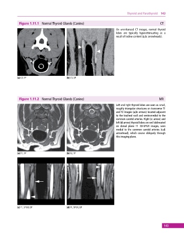
Figure 1.11.1 Normal Thyroid Glands (Canine) CT
On unenhanced CT images, normal thyroid
lobes are typically hyperattenuating as a
result of iodine content (a,b: arrowheads).
(a) CT, TP (b) CT, DP
Figure 1.11.2 Normal Thyroid Glands (Canine) MR
Left and right thyroid lobes are seen as small,
roughly triangular structures on transverse T1
and T2 images (a,b: arrows) located adjacent
to the tracheal wall and ventromedial to the
common carotid arteries. Right (c: arrow) and
left (d: arrow) thyroid lobes are well delineated
on dorsal plane T1 3D‐SPGR images, seen
medial to the common carotid arteries (c,d:
arrowhead), which course obliquely through
this imaging plane.
(a) T1, TP (b) T2, TP
(c) T1, SPGR, DP (d) T1, SPGR, DP
143