
Page 154 - Atlas of Small Animal CT and MRI
P. 154
144 Atlas of Small Animal CT and MRI
Figure 1.11.3 Hypothyroidism (Canine) CT
12y FS Weimaraner with documented
hypothyroidism. Image b is the same image
as a with line overlays showing the long‐
axis oblique planes depicted in c and d. The
thyroid lobes are smaller than expected and
are marginally hyperattenuating than adja-
cent soft tissues (a: arrows). The right (c:
arrows) and left (d: arrows) thyroid lobes are
easily delineated on the oblique images.
Both lobes are small, and margins are abnor-
mally lobular. Thyroid lobes viewed in long
axis are distinguished from the medial
retropharyngeal lymph nodes which can
appear similar but are more cranial and
(a) CT, TP (b) CT, TP
located lateral to the neurovascular bundle.
(C) CT, OP (d) CT, OP
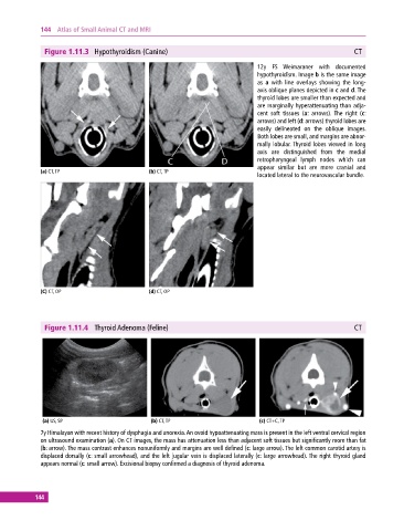
Figure 1.11.4 Thyroid Adenoma (Feline) CT
(a) US, SP (b) CT, TP (c) CT+C, TP
7y Himalayan with recent history of dysphagia and anorexia. An ovoid hypoattenuating mass is present in the left ventral cervical region
on ultrasound examination (a). On CT images, the mass has attenuation less than adjacent soft tissues but significantly more than fat
(b: arrow). The mass contrast enhances nonuniformly and margins are well defined (c: large arrow). The left common carotid artery is
displaced dorsally (c: small arrowhead), and the left jugular vein is displaced laterally (c: large arrowhead). The right thyroid gland
appears normal (c: small arrow). Excisional biopsy confirmed a diagnosis of thyroid adenoma.
144