
Page 158 - Atlas of Small Animal CT and MRI
P. 158
148 Atlas of Small Animal CT and MRI
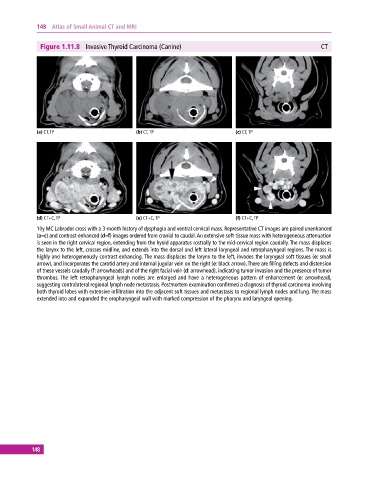
Figure 1.11.8 Invasive Thyroid Carcinoma (Canine) CT
(a) CT, TP (b) CT, TP (c) CT, TP
(d) CT+C, TP (e) CT+C, TP (f) CT+C, TP
10y MC Labrador cross with a 3‐month history of dysphagia and ventral cervical mass. Representative CT images are paired unenhanced
(a–c) and contrast‐enhanced (d–f) images ordered from cranial to caudal. An extensive soft‐tissue mass with heterogeneous attenuation
is seen in the right cervical region, extending from the hyoid apparatus rostrally to the mid‐cervical region caudally. The mass displaces
the larynx to the left, crosses midline, and extends into the dorsal and left lateral laryngeal and retropharyngeal regions. The mass is
highly and heterogeneously contrast enhancing. The mass displaces the larynx to the left, invades the laryngeal soft tissues (e: small
arrow), and incorporates the carotid artery and internal jugular vein on the right (e: black arrow). There are filling defects and distension
of these vessels caudally (f: arrowheads) and of the right facial vein (d: arrowhead), indicating tumor invasion and the presence of tumor
thrombus. The left retropharyngeal lymph nodes are enlarged and have a heterogeneous pattern of enhancement (e: arrowhead),
suggesting contralateral regional lymph node metastasis. Postmortem examination confirmed a diagnosis of thyroid carcinoma involving
both thyroid lobes with extensive infiltration into the adjacent soft tissues and metastasis to regional lymph nodes and lung. The mass
extended into and expanded the oropharyngeal wall with marked compression of the pharynx and laryngeal opening.
148