Page 161 - Atlas of Small Animal CT and MRI
P. 161

Thyroid and Parathyroid  151


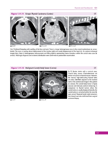
              Figure 1.11.11  Ectopic Thyroid Carcinoma (Canine)                                          CT


            (a) CT, TP                       (b) CT+C, TP                     (c) CT+C, SP
            10y F Shetland Sheepdog with swelling of the face and neck. There is a large, heterogeneous mass in the cranial mediastinum (a: arrow-
            heads). The mass is causing dorsal displacement of the trachea (a,b) and caudal displacement of the heart (c). On contrast‐enhanced
            images (b,c), there is heterogeneous enhancement and filling defects representing tumor thrombus within the cranial vena cava (b:
            arrows). Histologic diagnosis and anatomic distribution were confirmed on postmortem examination.


              Figure 1.11.12  Malignant Carotid Body Tumor (Canine)                                       CT

                                                                             9y FS Boston terrier with a cervical mass.
                                                                             Carotid  body  tumors  (Chemodectoma)  are
                                                                             similar in location to thyroid masses. However,
                                                                             the  normal,  high‐attenuating  thyroid  lobes
                                                                             are easily identified adjacent to the tracheal
                                                                             wall on the unenhanced image in this patient
                                                                             (a: arrows). The carotid body tumor is highly
                                                                             vascular and intensely contrast enhancing. In
                                                                             comparison to thyroid tumors where the
                                                                             carotid artery is usually displaced laterally, the
                                                                             carotid artery is contained within the mass (b:
                                                                             open arrow). Regional lymph node metastasis
                                                                             (not shown) was also confirmed from surgical
                                                                             excisional biopsy.
            (a) CT, TP                       (b) CT+C, TP


                                                                                                             151
   156   157   158   159   160   161   162   163   164   165   166