
Page 162 - Atlas of Small Animal CT and MRI
P. 162
152 Atlas of Small Animal CT and MRI
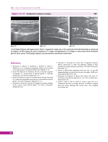
Figure 1.11.13 Parathyroid Carcinoma (Canine) MR
(a) US, SP (b) T1, SP (c) T2, SP
12y M Golden Retriever with hypercalcemia. There is a hypoechoic nodule seen in the cranial pole of the left thyroid lobe on ultrasound
(a: calipers). On MR imaging, the mass is isointense on T1 images and hyperintense on T2 images (c: white arrow) within the thyroid
gland (c: black arrows). The histologic diagnosis was documented on postmortem examination.
References 5. Taeymans O, Penninck DG, Peters RM. Comparison between
clinical, ultrasound, CT, MRI, and pathology findings in dogs
1. Taeymans O, Schwarz T, Duchateau L, Barberet V, Gielen I, presented for suspected thyroid carcinoma. Vet Radiol Ultrasound.
Haskins M, et al. Computed tomographic features of the normal 2012;54:61–70.
canine thyroid gland. Vet Radiol Ultrasound. 2008;49:13–19. 6. Jhaveri K, Shroff MM, Fatterpekar GM, Som PM. CT and MR
2. Drost WT, Mattoon JS, Weisbrode SE. Use of helical computed imaging findings associated with subacute thyroiditis. AJNR Am J
tomography for measurement of thyroid glands in clinically Neuroradiol. 2003;24:143–146.
normal cats. Am J Vet Res. 2006;67:467–471. 7. Hofmeister E, Kippenes H, Mealey KL, Cantor GH, Lohr CV.
3. Drost WT, Mattoon JS, Samii VF, Weisbrode SE, Hoshaw‐Woodard Functional cystic thyroid adenoma in a cat. J Am Vet Med Assoc.
SL. Computed tomographic densitometry of normal feline thyroid 2001;219:190–193.
glands. Vet Radiol Ultrasound. 2004;45:112–116. 8. Cakal E, Cakir E, Dilli A, Colak N, Unsal I, Aslan MS, et al.
4. Taeymans O, Dennis R, Saunders JH. Magnetic resonance imaging Parathyroid adenoma screening efficacies of different imaging
of the normal canine thyroid gland. Vet Radiol Ultrasound. tools and factors affecting the success rates. Clin Imaging.
2008;49:238–242. 2012;36:688–694.
152