
Page 167 - Atlas of Small Animal CT and MRI
P. 167
Ventricular System and Hydrocephalus 157
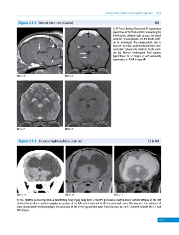
Figure 2.1.2 Normal Ventricles (Canine) MR
7y FS French Bulldog. The normal T1 hypointense
appearance of the third ventricle surrounding the
interthalamic adhesion (a,b: arrows), the lateral
ventricles (b: arrowheads), and the fourth ventri-
cle (a: arrowhead). The mesencephalic duct is
also seen as a thin, curvilinear, hypointense com-
munication between the third and fourth ventri-
cles (a). Normal cerebrospinal fluid appears
hyperintense on T2 images (c) and profoundly
hypointense on FLAIR images (d).
(a) T1, SP (b) T1, TP
(c) T2, TP (d) FL, TP
Figure 2.1.3 Ex vacuo Hydrocephalus (Canine) CT & MR
(a) CT, TP (b) T1, TP (c) T2, TP
4y MC Maltese recovering from a penetrating head injury (dog bite) 6 months previously. Posttraumatic cortical atrophy of the left
cerebral hemisphere results in passive expansion of the left lateral ventricle to fill the potential space. This dog also has evidence of
more generalized ventriculomegaly. Discontinuity of the overlying parietal bone from previous fracture is evident on both the CT and
MR images.
157