
Page 169 - Atlas of Small Animal CT and MRI
P. 169
Ventricular System and Hydrocephalus 159
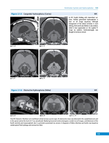
Figure 2.1.5 Congenital Hydrocephalus (Canine) MR
4y MC English Bulldog with intermittent sei-
zures. Marked generalized hydrocephalus is
seen on all image sequences. Although
enlargement of the lateral ventricles is most
striking, third ventricular dilation is also evident,
which is best appreciated on the sagittal T1
image (a: asterisk). Ventriculomegaly was
thought to be breed related.
(a) T1, SP (b) T1, TP
(c) T2, TP (d) FL, TP
Figure 2.1.6 Obstructive Hydrocephalus (Feline) MR
(a) T2, SP (b) T2, TP (c) T1+C, TP
7mo MC Domestic Shorthair with multifocal central nervous system signs. An obstructive mass was detected in the caudal brainstem and
in the spinal cord at the level of C1 (a: arrowhead). Marked generalized ventriculomegaly is evident on all images, and distension of the
fourth ventricle and mesencephalic duct is particularly prominent (a: arrows). A diagnosis of feline infectious peritonitis was based on
cerebrospinal fluid cytology and coronavirus titers.
159