
Page 168 - Atlas of Small Animal CT and MRI
P. 168
158 Atlas of Small Animal CT and MRI
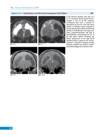
Figure 2.1.4 Hydrocephalus with Abnormal Cerebrospinal Fluid (Feline) MR
2y MC Domestic Shorthair with signs of a
C1–C5 myelopathy. Bilateral lateral ventricu-
lomegaly is seen on all MR sequences.
Cerebrospinal fluid (CSF) is uniformly T2
hyperintense (a), but CSF in the right lateral
ventricle is moderately intense compared to
the low‐intensity signal within the left lateral
ventricle on FLAIR (b) and T1 (c) images, indi-
cating compartmentalization and cells or
macromolecules contaminating the CSF in
the right lateral ventricle. Thickening and
intense enhancement of the right lateral
ependymal lining is evident on the contrast‐
enhanced T1 image (d). A diagnosis of feline
(a) T2, TP (b) FL, TP infectious peritonitis was based on cerebro-
spinal fluid cytology and coronavirus titers.
(c) T1, TP (d) T1+C, TP
158