
Page 160 - Atlas of Small Animal CT and MRI
P. 160
150 Atlas of Small Animal CT and MRI
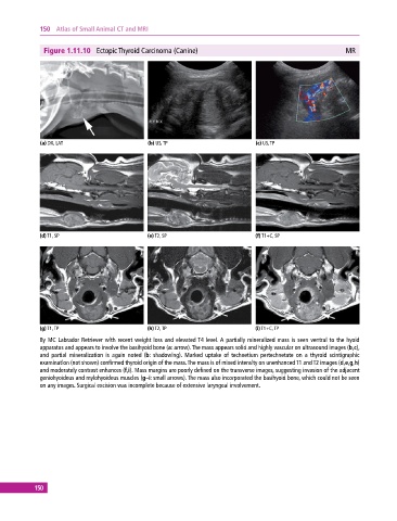
Figure 1.11.10 Ectopic Thyroid Carcinoma (Canine) MR
(a) DX, LAT (b) US, TP (c) US, TP
(d) T1, SP (e) T2, SP (f) T1+C, SP
(g) T1, TP (h) T2, TP (i) T1+C, TP
8y MC Labrador Retriever with recent weight loss and elevated T4 level. A partially mineralized mass is seen ventral to the hyoid
apparatus and appears to involve the basihyoid bone (a: arrow). The mass appears solid and highly vascular on ultrasound images (b,c),
and partial mineralization is again noted (b: shadowing). Marked uptake of technetium pertechnetate on a thyroid scintigraphic
examination (not shown) confirmed thyroid origin of the mass. The mass is of mixed intensity on unenhanced T1 and T2 images (d,e,g,h)
and moderately contrast enhances (f,i). Mass margins are poorly defined on the transverse images, suggesting invasion of the adjacent
geniohyoideus and mylohyoideus muscles (g–i: small arrows). The mass also incorporated the basihyoid bone, which could not be seen
on any images. Surgical excision was incomplete because of extensive laryngeal involvement.
150