
Page 157 - Atlas of Small Animal CT and MRI
P. 157
Thyroid and Parathyroid 147
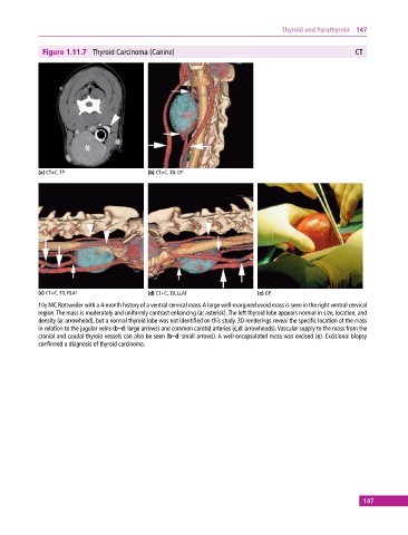
Figure 1.11.7 Thyroid Carcinoma (Canine) CT
(a) CT+C, TP (b) CT+C, 3D, OP
(c) CT+C, 3D, RLAT (d) CT+C, 3D, LLAT (e) GP
10y MC Rottweiler with a 4‐month history of a ventral cervical mass. A large well‐margined ovoid mass is seen in the right ventral cervical
region. The mass is moderately and uniformly contrast enhancing (a: asterisk). The left thyroid lobe appears normal in size, location, and
density (a: arrowhead), but a normal thyroid lobe was not identified on this study. 3D renderings reveal the specific location of the mass
in relation to the jugular veins (b–d: large arrows) and common carotid arteries (c,d: arrowheads). Vascular supply to the mass from the
cranial and caudal thyroid vessels can also be seen (b–d: small arrows). A well‐encapsulated mass was excised (e). Excisional biopsy
confirmed a diagnosis of thyroid carcinoma.
147