
Page 159 - Atlas of Small Animal CT and MRI
P. 159
Thyroid and Parathyroid 149
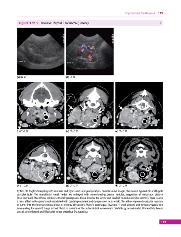
Figure 1.11.9 Invasive Thyroid Carcinoma (Canine) CT
(a) US, SP (b) US, SP
(c) CT+C, TP (d) CT+C, TP (e) CT+C, TP
(f) CT+C, TP (g) CT+C, TP (h) CT+C, TP
6y MC Old English Sheepdog with anorexia and right‐sided laryngeal paralysis. On ultrasound images, the mass is hypoechoic and highly
vascular (a,b). The mandibular lymph nodes are enlarged with nonenhancing central nodules, suggestive of metastatic disease
(c: arrowhead). The diffuse, contrast‐enhancing neoplastic tissue invades the larynx and cervical musculature (d,e: arrows). There is also
a mass effect in the spinal canal associated with cord displacement and compression (e: asterisk). This either represents vascular invasion
of tumor into the internal venous plexus or venous obstruction. There is esophageal invasion (f: small arrows) and tortuous vasculature
surrounding the mass (f: large arrow). There is invasion of the subvertebral musculature caudally (g: arrowheads). Unidentified tumor
vessels are enlarged and filled with tumor thrombus (h: asterisks).
149