
Page 156 - Atlas of Small Animal CT and MRI
P. 156
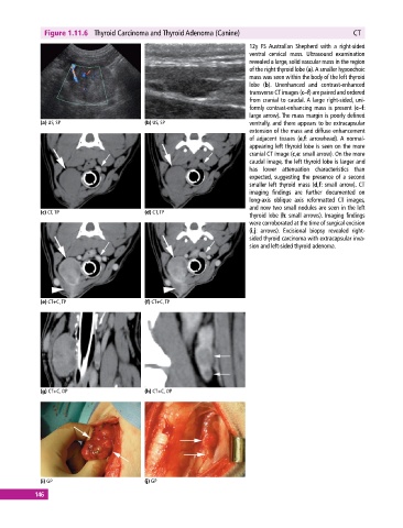
Figure 1.11.6  Thyroid Carcinoma and Thyroid Adenoma (Canine)                                CT

                                                                            12y FS  Australian Shepherd with a right‐sided
                                                                            ventral cervical mass. Ultrasound examination
                                                                            revealed a large, solid vascular mass in the region
                                                                            of the right thyroid lobe (a). A smaller hypoechoic
                                                                            mass was seen within the body of the left thyroid
                                                                            lobe (b). Unenhanced and contrast‐enhanced
                                                                            transverse CT images (c–f) are paired and ordered
                                                                            from cranial to caudal. A large right‐sided, uni-
                                                                            formly contrast‐enhancing mass is present (c–f:
                                                                            large arrow). The mass margin is poorly defined
             (a) US, SP                     (b) US, SP                      ventrally, and there appears to be extracapsular
                                                                            extension of the mass and diffuse enhancement
                                                                            of adjacent tissues (e,f: arrowhead). A normal‐
                                                                            appearing left thyroid lobe is seen on the more
                                                                            cranial CT image (c,e: small arrow). On the more
                                                                            caudal image, the left thyroid lobe is larger and
                                                                            has lower attenuation characteristics than
                                                                            expected, suggesting the presence of a second
                                                                            smaller left thyroid mass (d,f: small arrow). CT
                                                                            imaging findings are further documented on
                                                                            long‐axis  oblique  axis  reformatted  CT  images,
                                                                            and now two small nodules are seen in the left
             (c) CT, TP                     (d) CT, TP
                                                                            thyroid lobe (h: small arrows). Imaging findings
                                                                            were corroborated at the time of surgical excision
                                                                            (i,j: arrows). Excisional biopsy revealed right‐
                                                                            sided thyroid carcinoma with extracapsular inva-
                                                                            sion and left‐sided thyroid adenoma.


             (e) CT+C, TP                   (f) CT+C, TP


             (g) CT+C, OP                   (h) CT+C, OP


             (i) GP                         (j) GP
           146