
Page 149 - Atlas of Small Animal CT and MRI
P. 149
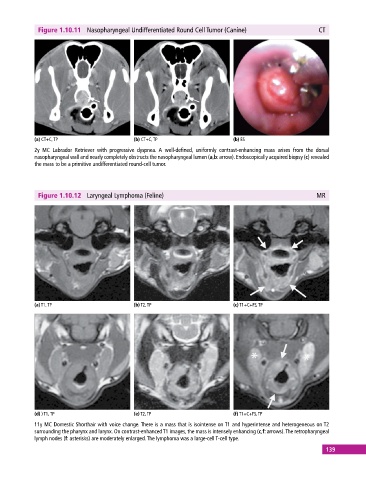
Figure 1.10.11  Nasopharyngeal Undifferentiated Round Cell Tumor (Canine)                   CT


            (a) CT+C, TP                     (b) CT+C, TP                     (b) ES
            2y MC Labrador Retriever with progressive dyspnea.  A well‐defined, uniformly contrast‐enhancing mass arises from the dorsal
              nasopharyngeal wall and nearly completely obstructs the nasopharyngeal lumen (a,b: arrow). Endoscopically acquired biopsy (c) revealed
            the mass to be a primitive undifferentiated round‐cell tumor.


              Figure 1.10.12  Laryngeal Lymphoma (Feline)                                                MR


            (a) T1, TP                       (b) T2, TP                       (c) T1+C+FS, TP


            (d) ) T1, TP                     (e) T2, TP                       (f) T1+C+FS, TP
            11y MC Domestic Shorthair with voice change. There is a mass that is isointense on T1 and hyperintense and heterogeneous on T2
              surrounding the pharynx and larynx. On contrast‐enhanced T1 images, the mass is intensely enhancing (c,f: arrows). The retropharyngeal
            lymph nodes (f: asterisks) are moderately enlarged. The lymphoma was a large‐cell T‐cell type.
                                                                                                             139