
Page 69 - Live-cellanalysis handbook
P. 69
Kinetic Antibody Internalization Assays
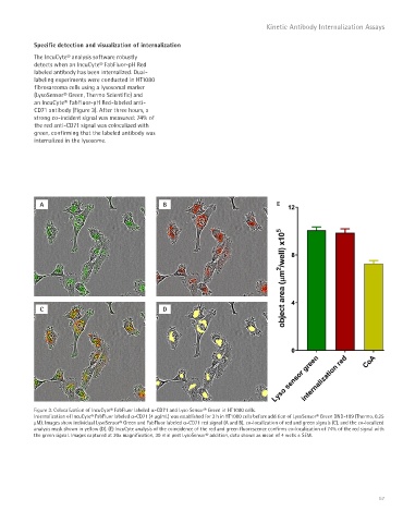
Specific detection and visualization of internalization
The IncuCyte® analysis software robustly
detects when an IncuCyte® FabFluor-pH Red
labeled antibody has been internalized. Dual-
labeling experiments were conducted in HT1080
fibrosarcoma cells using a lysosomal marker
(LysoSensor® Green, Thermo Scientific) and
an IncuCyte® FabFluor-pH Red-labeled anti-
CD71 antibody (Figure 3). After three hours, a
strong co-incident signal was measured: 74% of
the red anti-CD71 signal was colocalized with
green, confirming that the labeled antibody was
internalized in the lysosome.
A B E
C D
Figure 3. Colocalization of IncuCyte® FabFluor labeled a-CD71 and Lyso Sensor® Green in HT1080 cells.
Internalization of IncuCyte® FabFluor labeled a-CD71 (4 μg/mL) was established for 3 h in HT1080 cells before addition of LysoSensor® Green DND-189 (Thermo, 0.25
μM). Images show individual LysoSensor® Green and FabFluor labeled a-CD71 red signal (A and B), co-localization of red and green signals (C), and the co-localized
analysis mask shown in yellow (D). (E) IncuCyte analysis of the coincidence of the red and green fluorescence confirms co-localization of 74% of the red signal with
the green signal. Images captured at 20x magnification, 30 min post LysoSensor® addition, data shown as mean of 4 wells ± SEM.
67