
Page 72 - Live-cellanalysis handbook
P. 72
Live-Cell Analysis Handbook — Third Edition
Comparison of multiple test antibodies for high-throughput screening
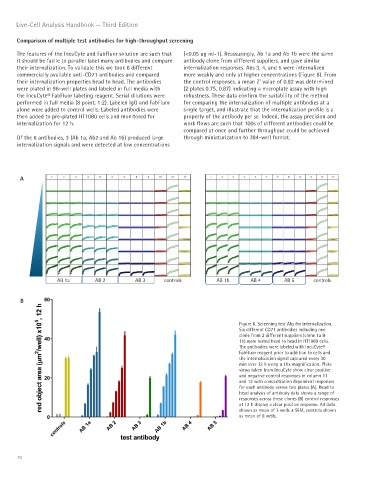
The features of the IncuCyte and FabFluor solution are such that (<0.05 ug ml-1). Reassuringly, Ab 1a and Ab 1b were the same
it should be facile to parallel label many antibodies and compare antibody clone from different suppliers, and gave similar
their internalization. To validate this we took 6 different internalization responses. Abs 3, 4, and 5 were internalized
commercially available anti-CD71 antibodies and compared more weakly and only at higher concentrations (Figure 8). From
their internalization properties head to head. The antibodies the control responses, a mean Z’ value of 0.82 was determined
were plated in 96-well plates and labeled in full media with (2 plates 0.75, 0.87) indicating a microplate assay with high
the IncuCyte® FabFluor labeling reagent. Serial dilutions were robustness. These data confirm the suitability of the method
performed in full media (8 point, 1:2). Labeled IgG and FabFluor for comparing the internalization of multiple antibodies at a
alone were added to control wells. Labeled antibodies were single target, and illustrate that the internalization profile is a
then added to pre-plated HT1080 cells and monitored for property of the antibody per se. Indeed, the assay precision and
internalization for 12 h. work flows are such that 100s of different antibodies could be
compared at once and further throughput could be achieved
Of the 6 antibodies, 3 (Ab 1a, Ab2 and Ab 1b) produced large through miniaturization to 384-well format.
internalization signals and were detected at low concentrations
A
AB 1a AB 2 AB 3 controls AB 1b AB 4 AB 5 controls
B
Figure 8. Screening test Abs for internalization.
Six different CD71 antibodies including one
clone from 2 different suppliers (clone 1a &
1b) were tested head to head in HT1080 cells.
The antibodies were labeled with IncuCyte®
FabFluor reagent prior to addition to cells and
the internalization signal captured every 30
min over 12 h using a 10x magnification. Plate
views taken from IncuCyte show clear positive
and negative control responses in column 11
and 12 with concentration dependent responses
for each antibody across two plates (A). Head to
head analysis of antibody data shows a range of
responses across these clones (B) control responses
at 12 h display a clear positive response. All data
shown as mean of 3 wells ± SEM, controls shown
as mean of 8 wells.
70