
Page 76 - Live-cellanalysis handbook
P. 76
Live-Cell Analysis Handbook — Third Edition
Sample Results
Quantitative measurements of
surface protein dynamics
Given the current explosion of checkpoint inhibitor cancer
therapies, assays that expedite further studies on the regulation
of immune-cell signaling pathways in tumors are an area of
significant need. The below example illustrates how dynamic
changes in cell surface checkpoint proteins can be quantified
in living cells in response to an inflammatory stimulus, using
Programmed Death Ligand-1 (PD-L1) as an archetype (Figure 2).
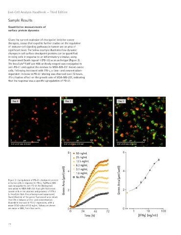
The IncuCyte® FabFluor-488 antibody reagent was conjugated to
anti-PD-L1 and applied the mixture to MDA-MB-231 breast cancer
cells. Following treatment with IFN-γ, a time- and concentration-
dependent increase in PD-L1 labeling was observed over 72 hours.
IFN-γ had no effect on the growth rate of MDA-MB-231, indicating
that the response was a specific upregulation of PD-L1.
Day 0 Day 1 Day 2
Figure 2: Upregulation of PD-L1 checkpoint protein
in tumor cells in response to IFN-γ. FabFluor-488
was conjugated to anti-PD-L1 Ab (BioLegend)
and added to MDA-MB-231 NucLight Red breast
cancer cells in the absence and presence of IFN-γ
(+ IncuCyte Opti-Green background suppressor).
Quantification of the green fluorescent area shows
that IFN-γ induces a time- and concentration-
dependent increase in PD-L1 expression, with a
mean EC50 value of 9.0 ng/mL. Values are shown
are mean ± SEM, from four wells.
74