
Page 75 - Live-cellanalysis handbook
P. 75
Live-cell Immunocytochemistry
Key advantages:
• Measure surface protein expression and distribution over • Visualize and quantify cell–cell interactions over time in complex
time using non-perturbing antibody labeling reagents in co-culture models, revealing insight into the interplay of cells.
physiologically relevant conditions.
• Significantly increase productivity compared to conventional
• Associate changes in surface protein expression with cell ICC by combining a rapid, single-step labeling protocol with
function and morphology to reveal informative, temporal automated acquisition and analysis. (Table 1).
changes in cell behavior.
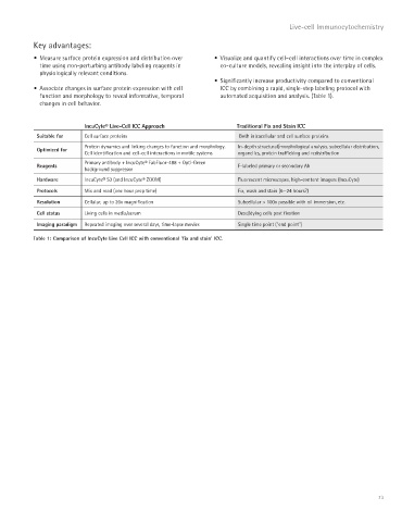
IncuCyte® Live-Cell ICC Approach Traditional Fix and Stain ICC
Suitable for Cell surface proteins Both intracellular and cell surface proteins
Protein dynamics and linking changes to function and morphology. In-depth structural/morphological analysis, subcellular distribution,
Optimized for
Cell identification and cell-cell interactions in motile systems organelles, protein trafficking and redistribution
Primary antibody + IncuCyte® FabFluor-488 + Opti-Green
Reagents F-labeled primary or secondary Ab
background suppressor
Hardware IncuCyte® S3 (and IncuCyte® ZOOM) Fluorescent microscopes, high-content imagers (IncuCyte)
Protocols Mix and read (one hour prep time) Fix, wash and stain (6—24 hours?)
Resolution Cellular, up to 20x magnification Subcellular > 100x possible with oil immersion, etc.
Cell status Living cells in media/serum Dead/dying cells post fixation
Imaging paradigm Repeated imaging over several days, time-lapse movies Single time point (“end point”)
Table 1: Comparison of IncuCyte Live Cell ICC with conventional ‘fix and stain’ ICC.
73