
Page 25 - Rapid Review of ECG Interpretation in Small Animal Practice, 2nd Edition
P. 25
Evaluation of the Electrocardiogram
HEART RATE • The regularity of the heartbeats: Are the RR
intervals regular or irregular?
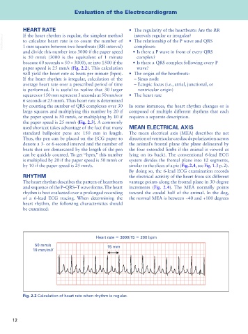
If the heart rhythm is regular, the simplest method
VetBooks.ir to calculate heart rate is to count the number of • The relationship of the P wave and QRS
complexes:
1 mm squares between two heartbeats (RR interval)
and divide this number into 3000 if the paper speed • Is there a P wave in front of every QRS
is 50 mm/s (3000 is the equivalent of 1 minute complex?
because 60 seconds × 50 = 3000), or into 1500 if the • Is there a QRS complex following every P
paper speed is 25 mm/s (Fig. 2.2). This calculation wave?
will yield the heart rate as beats per minute (bpm). • The origin of the heartbeats:
If the heart rhythm is irregular, calculation of the – Sinus node
average heart rate over a prescribed period of time – Ectopic focus (i.e., atrial, junctional, or
is performed. It is useful to realize that 30 larger ventricular origin)
squares or 150 mm represent 3 seconds at 50 mm/s or • The heart rate
6 seconds at 25 mm/s. Thus heart rate is determined
by counting the number of QRS complexes over 30 In some instances, the heart rhythm changes or is
large squares and multiplying this number by 20 if composed of multiple different rhythms that each
the paper speed is 50 mm/s, or multiplying by 10 if requires a separate description.
the paper speed is 25 mm/s (Fig. 2.3). A commonly
used shortcut takes advantage of the fact that many MEAN ELECTRICAL AXIS
standard ballpoint pens are 150 mm in length. The mean electrical axis (MEA) describes the net
Thus, the pen can be placed on the ECG paper to direction of ventricular cardiac depolarization across
denote a 3- or 6-second interval and the number of the animal’s frontal plane (the plane delineated by
beats that are demarcated by the length of the pen the four extended limbs if the animal is viewed as
can be quickly counted. To get “bpm,” this number lying on its back). The conventional 6-lead ECG
is multiplied by 20 if the paper speed is 50 mm/s or system divides the frontal plane into 12 segments,
by 10 if the paper speed is 25 mm/s. similar to the slices of a pie (Fig. 2.4, see Fig. 1.3 p. 2).
By doing so, the 6-lead ECG examination records
RHYTHM the electrical activity of the heart from six different
The heart rhythm describes the pattern of heartbeats vantage points along the frontal plane in 30-degree
and sequence of the P–QRS–T wave forms. The heart increments (Fig. 2.4). The MEA normally points
rhythm is best evaluated over a prolonged recording toward the caudal half of the animal. In the dog,
of a 6-lead ECG tracing. When determining the the normal MEA is between +40 and +100 degrees
heart rhythm, the following characteristics should
be examined:
Heart rate = 3000/15 = 200 bpm
50 mm/s 15 mm
10 mm/mV
Fig. 2.2 Calculation of heart rate when rhythm is regular.
12