
Page 89 - Rapid Review of ECG Interpretation in Small Animal Practice, 2nd Edition
P. 89
Answers 24, 25 ECG Cases
Answer 24
VetBooks.ir 1 ECG 24 shows sinus arrhythmia and criteria for atrial enlargement.
2 • The heart rate is ~100 bpm. The P waves are tall and peaked in leads I, II, and aVF. The amplitude of
the P wave in lead II is 0.5 mV (normal: <0.4 mV). A tall P wave is often referred to as “P pulmonale”
despite the fact that tall P waves can be detected in both cases of left or right atrial enlargement.
• At times, tall P waves are seen in patients with pulmonary disease, right ventricular hypertrophy, and
pulmonary hypertension (cor pulmonale).
Answer 25
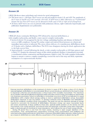
1 ECG 25 shows ventricular fibrillation (VF) followed by electrical defibrillation, a
wide complex tachycardia, and finally a more narrow complex tachycardia.
• Section A shows VF, recognized by the low voltage undulating baseline and absence of distinct P
waves or QRS complexes. During VF, cardiac output and arterial blood pressure are absent and
immediate intervention is indicated. The arrow shows delivery of a transthoracic defibrillation shock
of 50 Joules with a biphasic defibrillator. The ECG trace disappears during the shock application due
to the high current flow.
• Approximately 1 second following the shock, a wide complex tachycardia at 230 bpm appears and
is likely VT. Despite the abnormal nature of this initial postshock rhythm, it immediately provided
a coordinated cardiac contraction and blood pressure, which is vital. The wide complex tachycardia
begins to assume a narrow complex morphology toward the end of the strip and likely represents
resumption of a supraventricular rhythm.
25
A
1 s
External electrical defibrillation is the treatment of choice in cases of VF. In dogs, a dose of 2–3 J/kg for
external biphasic defibrillation is recommended. Direct current shocks are delivered to the heart in order
to depolarize uniformly and simultaneously a critical mass of the excitable myocardium. The objectives
are to interfere with all arrhythmia mechanisms and to allow the normal cardiac pacemakers to assume the
role of primary pacemaker. Failure to defibrillate or immediate recurrence of VF are an indication to repeat
defibrillation with a higher shock energy setting or more a optimized defibrillation set-up. Successful
defibrillation depends on two key factors: (1) duration between onset of fibrillation and defibrillation,
and (2) the metabolic condition of the myocardium. The goal is to use the minimum amount of energy
required to overcome the threshold of defibrillation. Excessive energy can cause myocardial injury
and worse arrhythmias. A large paddle-to-chest ratio, optimal paddle position on the thorax over the
myocardium, firm contact pressure, and shaved chest and adequate conduction (using either disposable
pads or electrode gel) all contribute to lower impedance, which allows the use of lower energy shocks.
During VF, blood flow to and from the heart ceases, resulting in rapid oxygen depletion of the
myocardium. Thus, cardiopulmonary resuscitation involving chest compressions and ventilation are
important while preparing the electric defibrillator or between failed defibrillation attempts. Antiarrhythmics
as a means to defibrillate are rarely effective, as perfusion of the myocardium is absent during VF.
76