
Page 151 - Fluid, Electrolyte, and Acid-Base Disorders in Small Animal Practice
P. 151
Disorders of Calcium: Hypercalcemia and Hypocalcemia 141
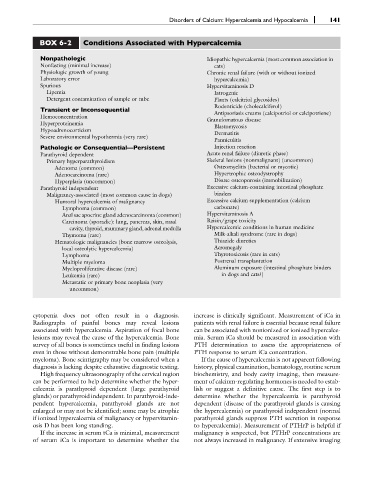
BOX 6-2 Conditions Associated with Hypercalcemia
Nonpathologic Idiopathic hypercalcemia (most common association in
Nonfasting (minimal increase) cats)
Physiologic growth of young Chronic renal failure (with or without ionized
Laboratory error hypercalcemia)
Spurious Hypervitaminosis D
Lipemia Iatrogenic
Detergent contamination of sample or tube Plants (calcitriol glycosides)
Transient or Inconsequential Rodenticide (cholecalciferol)
Antipsoriasis creams (calcipotriol or calcipotriene)
Hemoconcentration
Granulomatous disease
Hyperproteinemia
Blastomycosis
Hypoadrenocorticism
Dermatitis
Severe environmental hypothermia (very rare)
Panniculitis
Pathologic or Consequential—Persistent Injection reaction
Parathyroid dependent Acute renal failure (diuretic phase)
Primary hyperparathyroidism Skeletal lesions (nonmalignant) (uncommon)
Adenoma (common) Osteomyelitis (bacterial or mycotic)
Adenocarcinoma (rare) Hypertrophic osteodystrophy
Hyperplasia (uncommon) Disuse osteoporosis (immobilization)
Parathyroid independent Excessive calcium-containing intestinal phosphate
Malignancy-associated (most common cause in dogs) binders
Humoral hypercalcemia of malignancy Excessive calcium supplementation (calcium
Lymphoma (common) carbonate)
Anal sac apocrine gland adenocarcinoma (common) Hypervitaminosis A
Carcinoma (sporadic): lung, pancreas, skin, nasal Raisin/grape toxicity
cavity, thyroid, mammary gland, adrenal medulla Hypercalcemic conditions in human medicine
Thymoma (rare) Milk-alkali syndrome (rare in dogs)
Hematologic malignancies (bone marrow osteolysis, Thiazide diuretics
local osteolytic hypercalcemia) Acromegaly
Lymphoma Thyrotoxicosis (rare in cats)
Multiple myeloma Postrenal transplantation
Myeloproliferative disease (rare) Aluminum exposure (intestinal phosphate binders
Leukemia (rare) in dogs and cats?)
Metastatic or primary bone neoplasia (very
uncommon)
cytopenia does not often result in a diagnosis. increase is clinically significant. Measurement of iCa in
Radiographs of painful bones may reveal lesions patients with renal failure is essential because renal failure
associated with hypercalcemia. Aspiration of focal bone can be associated with nonionized or ionized hypercalce-
lesions may reveal the cause of the hypercalcemia. Bone mia. Serum iCa should be measured in association with
survey of all bones is sometimes useful in finding lesions PTH determination to assess the appropriateness of
even in those without demonstrable bone pain (multiple PTH response to serum iCa concentration.
myeloma). Bone scintigraphy may be considered when a If the cause of hypercalcemia is not apparent following
diagnosis is lacking despite exhaustive diagnostic testing. history, physical examination, hematology, routine serum
High frequency ultrasonography of the cervical region biochemistry, and body cavity imaging, then measure-
can be performed to help determine whether the hyper- ment of calcium-regulating hormones is needed to estab-
calcemia is parathyroid dependent (large parathyroid lish or suggest a definitive cause. The first step is to
glands) or parathyroid independent. In parathyroid-inde- determine whether the hypercalcemia is parathyroid
pendent hypercalcemia, parathyroid glands are not dependent (disease of the parathyroid glands is causing
enlarged or may not be identified; some may be atrophic the hypercalcemia) or parathyroid independent (normal
if ionized hypercalcemia of malignancy or hypervitamin- parathyroid glands suppress PTH secretion in response
osis D has been long standing. to hypercalcemia). Measurement of PTHrP is helpful if
If the increase in serum tCa is minimal, measurement malignancy is suspected, but PTHrP concentrations are
of serum iCa is important to determine whether the not always increased in malignancy. If extensive imaging