
Page 147 - Fluid, Electrolyte, and Acid-Base Disorders in Small Animal Practice
P. 147
Disorders of Calcium: Hypercalcemia and Hypocalcemia 137
above normal often have adverse pathophysiologic or during rapid infusion of calcium-containing fluids.
consequences. Hypercalcemia represents a clinically Dogs with similar magnitudes of hypercalcemia may dis-
relevant increase above an individual animal’s own nor- play minimal clinical signs when hypercalcemia has devel-
mal serum calcium concentration, usually defined as a oped gradually. Regardless of the rate of increase in serum
fasting serum tCa concentration greater than 12.0 mg/ calcium concentration, clinical signs become more severe
dL in dogs or greater than 11.0 mg/dL in cats. Ionized as the magnitude of hypercalcemia increases. Serum tCa
calcium measurements can provide greater sensitivity and concentrations of 12.0 to 14.0 mg/dL may not be
specificity for the diagnosis of some hypercalcemic associated with severe clinical signs, but most animals
disorders. A serum iCa concentration greater than with concentrations greater than 15.0 mg/dL show sys-
6.0 mg/dL (1.5 mmol/L) in dogs and greater than temic signs. Dogs with serum calcium concentrations
5.7 mg/dL (1.4 mmol/L) in cats constitutes ionized greater than 18 mg/dL are often severely ill, and
hypercalcemia. concentrations greater than 20 mg/dL may constitute
a life-threatening crisis. Exceptions do occur, however,
TOXICITY OF HYPERCALCEMIA AND and some dogs are severely affected by mild hypercalce-
CLINICAL SIGNS mia, whereas others are relatively unaffected by severe
Excessive calcium ions are toxic to cells, 462 and increased hypercalcemia. Clinical signs and histopathologic
serum iCa concentration decreases cellular function by changes are more likely to develop the longer hypercalce-
causing alterations in cell membrane permeability and cell mia has been present, regardless of its magnitude. Pro-
membrane calcium pump activity. Increased intracellular gressive hypercalcemia may also contribute to the
iCa content can ultimately result in cell death caused by severity of clinical signs, as occurs in animals with malig-
deranged cellular function and reduced energy produc- nant neoplasia or hypervitaminosis D related to rat bait
tion. Although all tissues may be subject to the dangerous ingestion.
effects of hypercalcemia, effects on the central nervous Changes in serum sodium and potassium
system, gastrointestinal tract, heart, and kidneys are of concentrations can magnify the clinical signs of hypercal-
most importance clinically. cemia by their effects on cell membrane excitability, par-
Polydipsia, polyuria, anorexia, lethargy, and weakness ticularly in nerve and muscle (see Chapter 5). Acidosis
are the most common clinical signs in dogs with hypercal- increases the proportion of serum calcium that is ionized,
cemia, 113,178 but individual animals often display remark- worsening clinical signs, whereas alkalosis lessens toxicity
able differences in clinical signs despite similar and clinical signs by decreasing the proportion of calcium
magnitudes of hypercalcemia. The severity of clinical that is ionized.
signs and development of lesions of hypercalcemia Mineralization of soft tissues (especially the heart and
depend not only on the magnitude of hypercalcemia kidneys) is an important complication of hypercalcemia.
but also on its rate of development and duration. Simul- The serum phosphorus concentration at the time hyper-
taneous disturbances in other electrolyte concentrations calcemia develops is important in determining the extent
and in acid-base balance, as well as organ dysfunction sec- of soft tissue mineralization. Soft tissue mineralization is
ondary to hypercalcemia, all contribute to clinical signs, most severe when the product of calcium (mg/dL) times
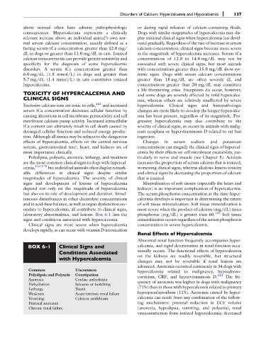
laboratory abnormalities, and lesions. Box 6-1 lists the phosphorus (mg/dL) is greater than 60. 115 Soft tissue
signs and conditions associated with hypercalcemia. mineralization occurs regardless of the serum phosphorus
Clinical signs are most severe when hypercalcemia concentration in severe hypercalcemia.
develops rapidly, as can occur with vitamin D intoxication
Renal Effects of Hypercalcemia
Abnormal renal function frequently accompanies hyper-
BOX 6-1 Clinical Signs and calcemia, and rapid deterioration in renal function occa-
Conditions Associated sionally occurs. The functional effects of hypercalcemia
with Hypercalcemia on the kidneys are readily reversible, but structural
changes may not be reversible if renal lesions are
advanced. Azotemia occurred commonly in 34 dogs with
Common Uncommon hypercalcemia related to malignancy, hypoadreno-
Polydipsia and Polyuria Constipation corticism, CRF, and hypervitaminosis D. 314 The fre-
Anorexia Cardiac arrhythmia quency of azotemia was higher in dogs with malignancy
Dehydration Seizures or twitching
(71%) than in those with hypercalcemia related to primary
Lethargy Death
hyperparathyroidism (11%). Azotemia caused by hyper-
Weakness Acute intrinsic renal failure
Vomiting Calcium urolithiasis calcemia can result from any combination of the follow-
Prerenal azotemia ing mechanisms: prerenal reduction in ECF volume
Chronic renal failure (anorexia, hypodipsia, vomiting, and polyuria); renal
vasoconstriction from ionized hypercalcemia; decreased