
Page 1099 - The Toxicology of Fishes
P. 1099
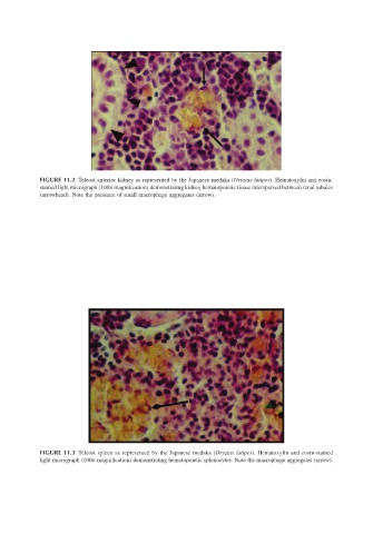
FIGURE 11.2 Teleost anterior kidney as represented by the Japanese medaka (Oryzias latipes). Hematoxylin and eosin-
                       stained light micrograph (100× magnification) demonstrating kidney hematopoietic tissue interspersed between renal tubules
                       (arrowhead). Note the presence of small macrophage aggregates (arrow).


                       FIGURE 11.3 Teleost spleen as represented by the Japanese medaka (Oryzias latipes). Hematoxylin and eosin-stained
                       light micrograph (100× magnification) demonstrating hematopoietic splenocytes. Note the macrophage aggregates (arrow).