
Page 1098 - The Toxicology of Fishes
P. 1098
(A) 100 µm (B) 100 µm
(C) 100 µm (D) 100 µm
FIGURE 7.16 In vivo imaging of tumor formation in STII medaka (brightfield microscopy). Neoplastic response following
early life stage exposure of STII medaka to the reference hepatocarcinogen diethylnitrosamine (DEN). Aqueous bath
exposure was of 24-hour duration at 100 ppm DEN. Fish were exposed as 14-day-old hatchlings and followed serially in
a noninvasive manner for 10 months. At 10 months after exposure, a subset of cohorts developed hepatic tumors (green
arrowhead). (A, C) In vivo imaging (brightfield) of hepatic tumor formation (green arrowheads) in DEN-exposed medaka;
shown is an enlargement of the total liver mass, approximately 18% of the total body length. After anesthesia, the liver was
removed and processed for histopathology. Hepatic neoplasm showed mixed hepatocellular (B) and cholangiocellular (D)
carcinomas. Focus of biliary hyperplasia was seen in the same liver; here, a single layer of biliary epithelium lines large
cystic spaces in the liver (D). The opaque white tissue in the brightfield images (A and B) is ovary; the gut occupies caudal
most region of the abdominal cavity.
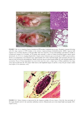
FIGURE 11.1 Teleost thymus as represented by the Japanese medaka (Oryzias latipes). Note the close proximity of
the organ (dashed circle) to the opercular epithelium (arrows). Hematoxylin and eosin-stained light micrograph (100×
magnification).