
Page 1095 - The Toxicology of Fishes
P. 1095
Bd
S
V
(A) 20 µm (B) 20 µm
Bd
S
S
(C) 20 µm (D) 20 µm
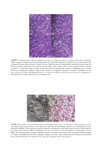
FIGURE 7.5 High-resolution light micrographs of medaka liver showing elements of hepatic architecture. (A) Hepatic
tubules comprised of hepatocytes and biliary epithelial cells are partially separated by sinusoids (S). No lobular architecture
is apparent. Rounded white structures are lipid vacuoles. Lipid was removed during alcohol dehydration in processing. (B)
Hepatic sinusoids contain nucleated red blood cells (top right of field). Larger venule (V) has no arterial or biliary structure
associated. (C) This field shows sinusoids (S) between which are found hepatic tubules in longitudinal array. Note the
double row of hepatocytes making a single tubule. Tubules are incompletely separated by sinusoids. (D) Region near the
hilus of the liver is shown. Intrahepatic bile ducts of varying sizes (Bd) are shown. Note the difference in staining and in
nuclear profiles in biliary epithelial cells vesus hepatocytes.
BPD
BPDC
(A) 2 µm (B) 10 µm
FIGURE 7.8 Canaliculi and transitional zones of intrahepatic biliary system are illustrated. (A) Transmission electron
micrograph of liver shows portions of two hepatocytes and one transitional biliary epithelial cell, termed bile preductular
epithelial cell (BPDC). This cell shares junctional complexes with hepatocytes and forms portion of wall of bile passageway
now termed bile preductule (BPD; a transitional zone). Note the absence of microvilli supplied to bile passageway by the
BPDC. (B) Light micrograph of paraffin-embedded sturgeon liver. Hepatocytes of this species contain little stainable material
in cytoplasm with H&E stain. Note the eosinophilic margins of the cells at bile canaliculi and their extensions toward tubule
lumen (black arrowheads). We seldom see bile canaliculi staining this well in other species.