
Page 1093 - The Toxicology of Fishes
P. 1093
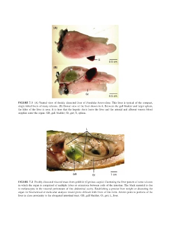
(A)


                                                                      S
                                                                            0.5 cm
                                           (B)      GB
                                                                      S


                                                              Gt
                                                                            0.5 cm
                       FIGURE 7.1 (A) Ventral view of freshly dissected liver of Fundulus heteroclitus. This liver is typical of the compact,
                       single-lobed livers of many teleosts. (B) Dorsal view of the liver shown in A. Between the gall bladder and larger spleen,
                       the hilus of the liver is seen. It is here that the hepatic ducts leave the liver and the arterial and afferent venous blood
                       supplies enter the organ. GB, gall bladder; Gt, gut; S, spleen.


                                                               L


                                                       GB         Gt         1 cm

                       FIGURE 7.2 Freshly dissected visceral mass from goldfish (Cyprinus carpio) illustrating the liver pattern of some teleosts
                       in which the organ is comprised of multiple lobes or extensions between coils of the intestine. The black material is due
                       to melanocytes in the visceral peritoneum of this abdominal cavity. Establishing a precise liver weight or dissecting the
                       organ for biochemical or molecular analyses would prove difficult with livers of this form. Arrows point to portions of the
                       liver in close proximity to the elongated intestinal tract. GB, gall bladder; Gt, gut; L, liver.