
Page 1097 - The Toxicology of Fishes
P. 1097
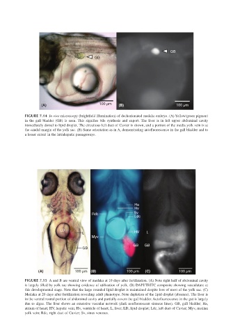
GB
                                                     GB


                              (A)                      100 µm  (B)                      100 µm

                       FIGURE 7.14 In vivo microscopy (brightfield illumination) of dechorionated medaka embryo. (A) Yellow/green pigment
                       in the gall bladder (GB) is seen.  This signifies bile synthesis and export.  The liver is in left upper abdominal cavity
                       immediately dorsal to lipid droplet. The circuitous left duct of Cuvier is shown, and a portion of the media yolk vein is at
                       the caudal margin of the yolk sac. (B) Same orientation as in A, demonstrating autofluorescence in the gall bladder and to
                       a lesser extent in the intrahepatic passageways.


                                                                      Ha
                                                                      Hv
                                                    Rdc               Sv
                                                                      Ldc

                                      LD
                                               L                      HV   L
                                                    Myv
                                                                     GB   GB
                                               GB


                            (A)             100 µm (B)             100 µm  (C)           100 µm

                       FIGURE 7.15 A and B are ventral view of medaka at 10 days after fertilization. (A) Note right half of abdominal cavity
                       is largely filled by yolk sac showing evidence of utilization of yolk. (B) DAPI/TRITC composite showing vasculature at
                       this developmental stage. Note that the large rounded lipid droplet is maintained despite loss of most of the yolk sac. (C)
                       Medaka at 20 days after fertilization revealing adult phenotype. Note depletion of the lipid droplet (absence). The liver is
                       in the ventral rostral portion of abdominal cavity and partially covers the gall bladder. Autofluorescence in the gut is largely
                       due to algae. The liver shows an extensive vascular network (dark nonfluorescent sinuous lines). GB, gall bladder; Ha,
                       atrium of heart; HV, hepatic vein; Hv, ventricle of heart; L, liver; LD, lipid droplet; Ldc, left duct of Cuvier; Myv, median
                       yolk vein; Rdc, right duct of Cuvier; Sv, sinus venosus.