Page 572 - The Toxicology of Fishes
P. 572

552                                                        The Toxicology of Fishes


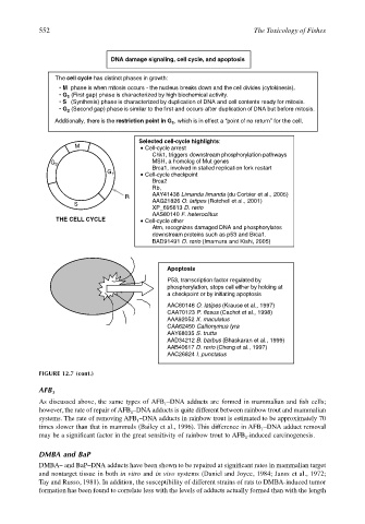
                                          DNA damage signaling, cell cycle, and apoptosis


                           The cell cycle has distinct phases in growth:
                            • M  phase is when mitosis occurs - the nucleus breaks down and the cell divides (cytokinesis).
                            • G 1  (First gap) phase is characterized by high biochemical activity.
                            • S   (Synthesis) phase is characterized by duplication of DNA and cell contents ready for mitosis.

                            • G  (Second gap) phase is similar to the first and occurs after duplication of DNA but before mitosis.
                              2
                           Additionally, there is the restriction point in G , which is in effect a “point of no returnʼʼ for the cell.
                                                           1
                                                  Selected cell-cycle highlights:
                                 M                  Cell-cycle arrest
                                                      Chk1, triggers downstream phosphorylation pathways
                          G 2                         MSH, a homolog of Mut genes
                                                      Brca1, involved in stalled replication fork restart
                                         G 1        Cell-cycle checkpoint
                                                      Brca2
                                                      Rb,
                                              R       AAY41438 Limanda limanda (du Corbier et al., 2005)
                                S                     AAG21826 O. latipes (Rotchell et al., 2001)
                                                      XP_695613 D. rerio
                                                      AAS80140 F. heteroclitus
                           THE CELL CYCLE           Cell-cycle other
                                                      Atm, recognizes damaged DNA and phosphorylates
                                                      downstream proteins such as p53 and Brca1.
                                                      BAD91491 D. rerio (Imamura and Kishi, 2005)


                                                          Apoptosis

                                                          P53, transcription factor regulated by
                                                          phosphorylation, stops cell either by holding at
                                                          a checkpoint or by initiating apoptosis
                                                          AAC60146 O. latipes (Krause et al., 1997)
                                                          CAA70123 P. flesus (Cachot et al., 1998)
                                                          AAA92052 X. maculatus
                                                          CAA62450 Callionymus lyra
                                                          AAY68035 S. trutta
                                                          AAD34212 B. barbus (Bhaskaran et al., 1999)
                                                          AAB40617 D. rerio (Cheng et al., 1997)
                                                          AAC26824 I. punctatus

                       FIGURE 12.7 (cont.)

                       AFB 1
                       As discussed above, the same types of AFB –DNA adducts are formed in mammalian and fish cells;
                                                         1
                       however, the rate of repair of AFB –DNA adducts is quite different between rainbow trout and mammalian
                                                1
                       systems. The rate of removing AFB –DNA adducts in rainbow trout is estimated to be approximately 70
                                                  1
                       times slower than that in mammals (Bailey et al., 1996). This difference in AFB –DNA adduct removal
                                                                                    1
                       may be a significant factor in the great sensitivity of rainbow trout to AFB -induced carcinogenesis.
                                                                                1
                       DMBA and BaP
                       DMBA– and BaP–DNA adducts have been shown to be repaired at significant rates in mammalian target
                       and nontarget tissue in both in vitro and in vivo systems (Daniel and Joyce, 1984; Janss et al., 1972;
                       Tay and Russo, 1981). In addition, the susceptibility of different strains of rats to DMBA-induced tumor
                       formation has been found to correlate less with the levels of adducts actually formed than with the length
   567   568   569   570   571   572   573   574   575   576   577