
Page 639 - The Toxicology of Fishes
P. 639
Toxicity Resistance 619
100 KCF1
AWF1
80 AWF2
% Survival 60
40
20
0
1:1 5:1 10:1
AW Sediment Dilution
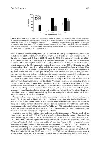
FIGURE 13.13 Survival of Atlantic Wood (creosote-contaminated site) and reference-site (King Creek) mummichog
progeny exposed to Atlantic Wood sediment. Progeny were hatched and raised in a clean laboratory environment and
exposed for 14 days to Atlantic Wood sediment diluted (1:1, 5:1, 10:1) with reference-site sediment. AWF1, first-generation
progeny of Atlantic Wood adults; AWF2, second-generation progeny of Atlantic Wood adults; KCF1, first-generation King
Creek progeny. Exposure to 1:1 dilutions resulted in 100% mortality of KCF1 and AWF2. (From Meyer, J.N. and Di Giulio,
R.T., Ecol. Appl., 13, 490–503, 2003. With permission.)
reared F embryos and larvae (Meyer et al., 2002); however, inducibility was regained as Atlantic Wood
1
F matured into adults. Further, inducibility was observed in all life stages of later generations raised in
1
the laboratory (Meyer and Di Giulio, 2002; Meyer et al., 2002). This pattern of reversible heritability
of the CYP1A phenotype was not explained by maternal effect (Meyer et al., 2002), altered transcription
of known CYP1A transcription factors (AHH, AhRR) (Meyer et al., 2003a), or hypermethylation of
specific cytosines in the CYP1A promoter region (Timme-Laragy et al., 2005). Differential molecular
techniques have also been used to explore pollution tolerance in mummichog from the Atlantic Wood
site (Meyer et al., 2005). Following laboratory exposures to sediment from the Atlantic Wood site of
fish from tolerant and local sensitive populations, individual variation was high but many sequences
were expressed in a sex- and/or population-specific manner, including (potentially) novel genes and
those not thought previously to be associated with AhR responsiveness (Meyer et al., 2005).
Exposure to Atlantic Wood sediments caused increases in many of the antioxidant defenses tested in
laboratory-raised mummichog from both the Atlantic Wood and a reference-site population (Meyer et
al., 2003b). In that study, Atlantic Wood mummichog were more resistant to the toxicity of a model
prooxidant than were reference-site fish, and they exhibited elevated levels of several antioxidant defenses
in the absence of any chemical exposure. Bacanskas et al. (2004) also noted seasonal and sex-specific
responses to prooxidants in pollutant-tolerant and -sensitive mummichog from Virginia residence sites
after collection and following laboratory holding. These results suggest that elevated antioxidant defenses
might contribute to the resistant phenotype.
Van Veld and coworkers have investigated the relationship between toxicity resistance and cancer in
Atlantic Wood mummichog. These fish exhibit altered expression of proteins involved in biotransfor-
mation and efflux in a pattern similar to that observed in multidrug-resistant tumors and cancer cell
lines; for example, immunoblot analysis indicated reduced expression of CYP1A in hepatocellular
carcinoma and in the foci of cellular alteration (Van Veld et al., 1992). Immunohistochemical examination
of liver sections revealed similar low levels of CYP1A in hepatocellular carcinoma, exocrine pancreatic
tissue, bile ducts, and cholangiocellular proliferative lesions.
Similarities between drug-resistant tumor cells and xenobiotic resistance in Atlantic Wood fish are
also suggested by studies with GST. GST activity was elevated approximately fivefold in liver of Atlantic
Wood mummichog compared to that of reference-site fish (Armknecht et al., 1996; Van Veld et al.,
1991). Monoclonal antibodies against mummichog GST indicated a sixfold elevation of GST in the liver
of Atlantic Wood fish relative to that measured in reference fish (Figure 13.14). Sequence information
on the elevated GST suggests a relationship to theta-like GST (Van Veld et al., 2005) described in
mammals (Mainwaring et al., 1996) and in fish (Gallagher et al., 1999; Leaver et al., 1993).
In addition to alterations in phase I and II enzymes, Pgp transporters were elevated in liver of Atlantic
Wood fish relative to reference fish (Cooper et al., 1996, 1999a,b) and were intensely overexpressed in
hepatic tumors (Figure 13.15). Immunohistochemical staining of mummichog liver demonstrated this