Page 333 - Feline Cardiology
P. 333
344  Section J: Pericardial Diseases


                A


      Pericardial Diseases


                                                                              B

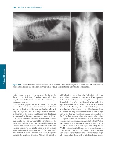
              Figure 22.1.  Lateral	(A)	and	VD	(B)	radiographs	from	a	cat	with	PPDH.	Note	the	severely	enlarged	cardiac	silhouette	with	overlap	of
              the	caudal	heart	border	and	diaphragm	and	the	presence	of	bowel	loops	containing	gas	within	the	pericardial	sac.


              major  organ  herniation  is  present.  Similarly,  the   midabdominal organs from the abdominal cavity may
              abdomen  may  feel  “empty.”  Other  congenital  defects   be noted and they may be visualized within the pericar-
              may also be noted such as sternebral abnormalities (i.e.,   dial sac. Echocardiography is a straightforward diagnos-
              pectus excavatum).                                 tic modality to confirm the diagnosis when abdominal
                 Electrocardiography may show reduced QRS ampli-  organs are visible within the pericardium of affected cats
              tude and/or axis deviation due to herniated abdominal   (Figure  22.2).  An  important  differential  diagnosis  is
              contents and shifted cardiac position. Radiographs typi-  consolidation of the accessory lung lobe, because hepa-
              cally are diagnostic, with an enlarged cardiac silhouette   tization of the lung mimics herniated liver tissue adja-
              and overlap of the caudal heart border and diaphragm   cent to the heart; a radiologist should be consulted to
              when organ herniation is moderate or extensive (Figure   clarify the diagnosis on radiographs if uncertainty exists.
              22.1). When  only  the  omentum  is  herniated,  thoracic   Surgical correction is warranted if clinical signs are
              radiographs  may  be  unremarkable.  Persistence  of  the   present, since the prognosis is excellent if the PPDH is
              dorsal mesothelial remnant, a meniscus-like connection   uncomplicated and repaired. In one study, 60% of the
              between  the  caudal  cardiac  silhouette  and  the  dia-  66  PPDH  cases  had  clinical  signs  secondary  to  the
              phragm,  ventral  to  the  caudal  vena  cava  on  a  lateral   PPDH, which prompted the owner to present the cat to
              radiograph, strongly suggests PPDH (O’Sullivan 2007).   a  veterinarian  (Reimer  et  al.  2004).  Twenty-nine  cats
              With herniation of one or more liver lobes, the gastric   were treated conservatively and 37 were treated surgi-
              axis  may  be  displaced  cranially. Absence  of  cranial  or   cally (most often those with overt clinical signs related

