Page 336 - Feline Cardiology
P. 336
Chapter 22: Pericardial Effusion and Other Disorders  347


                                                                 pericardiocentesis from the left or right precordium, but
                                                                 the ideal location can be best assessed via echocardiog-
                                                                 raphy (which allows one to choose the space with the
                                                                 largest fluid pocket). The patient should be restrained in
                                                                 lateral recumbency and the intercostal space should be
                                      LV
                                                                 blocked with 1 cc lidocaine using a 25–27 gauge needle.
                                            LA                   Some cats may need sedation in addition to the local
                                                                 lidocaine  block.  The  catheter/butterfly  should  be
                                     PE
                                                                 attached to a 3-way stopcock as described for thoraco-
                                                                 centesis  (see  discussion  regarding  pleural  effusion
                                                                 removal in Chapter 3). An electrocardiogram should be
                                                                 placed to evaluate for new ventricular arrhythmias that
                                                                 may occur if the ventricle is contacted. Complications
                                                                 secondary to pericardiocentesis are rare, but they include
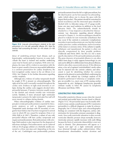
              Figure  22.4.  Long-axis	 echocardiogram	 obtained	 at	 the	 right	  cardiac puncture, cardiac arrhythmias, and/or laceration
              parasternum	 of	 a	 cat	 with	 pericardial	 effusion	 (PE).	 Note	 the	  of the tumor or coronary artery. If the catheter is rapidly
              anechoic	fluid	surrounding	the	heart.	LA	=	left	atrium;	LV	=	left	  withdrawn  and  repositioned,  the  patient  is  often  not
              ventricle.
                                                                 clinically  compromised  by  these  possible  problems
                                                                 (Gidlewski and Petrie 2005). See Box 22.1 for a more
              dence  of  underlying  primary  heart  disease,  such  as   specific description of the pericardiocentesis procedure.
              hypertrophic  cardiomyopathy;  however, in  some indi-  The gross character of pericardial effusion in cats often
              viduals  the  heart  is  normal  and  another  underlying   differs from dogs; it rarely appears hemorrhagic in cats
              cause may be found, such as neoplasia. With some neo-  and can be difficult to differentiate from pleural effusion,
              plasms, the mass will be evident in association with the   which is also often concurrently present. If this dilemma
              heart or great vessel(s); however, myocardial infiltration   arises, a few ml of agitated (microbubbled) sterile saline
              is  a  common  presentation  for  cardiac  lymphoma,  the   may  be  injected  into  the  centesis  catheter,  and  ultra-
              best-recognized cardiac tumor in the cat (Meurs et al.   sound  is  used  for  identifying  whether  the  contrast
              1994). See Chapter 16 for further discussion regarding   appears in the pleural or pericardial fluid, confirming the   Pericardial Diseases
              cardiac neoplasia.                                 location  of  the  catheter  tip.  Cytologic  analysis  of  PE
                 Although rare, evidence of cardiac tamponade should   should be performed if infectious or neoplastic etiolo-
              be  sought  if  PE  is  present  on  echocardiography.  The   gies are possible (i.e., most cases of PE causing cardiac
              right atrial free wall is normally rounded throughout the   tamponade  in  cats).  Malignant  lymphocytes  generally
              cardiac cycle. Evidence of right atrial inversion or col-  exfoliate  readily  in  feline  PE  caused  by  lymphoma
              lapse  during  the  cardiac  cycle  suggests  elevated  intra-  (Brummer and Moïse 1989).
              pericardial pressure. If present, inversion usually occurs
              in  late  diastole  and  extends  variably  into  ventricular   CONSTRICTIVE PERICARDITIS
              systole.  Similarly,  if  more  advanced  right  ventricular
              tamponade is present, the right ventricular free wall will   Pericardial constrictive disease occurs when the visceral
              move inward or collapse during diastole.           and parietal pericardial layers become fused or fibrotic
                 When  echocardiographic  evidence  of  cardiac  tam-  (Figure 22.5). The pericardial space may be totally oblit-
              ponade is noted, pericardiocentesis is essential for thera-  erated or may contain a small amount of PE. Constrictive
              peutic  purposes  as  well  as  diagnostic  purposes.   pericarditis is a difficult diagnosis to make by echocar-
              Tamponade rarely develops with PE secondary to con-  diography,  but  if  clinical  signs  and  echocardiographic
              gestive heart failure, but it has been recognized in several   findings are consistent with the diagnosis, cardiac cath-
              cases in which PE was caused by neoplasia (Zoia et al.   eterization for intracardiac pressure assessment is diag-
              2004; Hall et al. 2007). Therefore, a subset of cats with   nostic. Since early ventricular relaxation is often normal,
              pericardial  effusion  will  have  cardiac  tamponade  and   ventricular diastolic pressure drops normally; however,
              require pericardiocentesis. Pericar diocentesis with cyto-  ventricular pressure rapidly increases with early filling
              logic fluid analysis is also important for the diagnosis of   because the limit of ventricular distensibility is met very
              infectious etiologies. Pericar diocentesis can be achieved   early  in  diastole.  This  phenomenon  will  appear  as  a
              with an 18–20 gauge catheter or a butterfly needle con-  “square  root  sign”  on  a  right  ventricular  pressure
              nected to a three-way stop cock (depending on the size   trace  obtained  through  cardiac  catheterization.  Other
              of  the  cat).  Cardiologists  may  preferentially  perform   hemodynamic  abnormalities  include  elevation  and

