Page 335 - Feline Cardiology
P. 335
346  Section J: Pericardial Diseases


              primary cardiac rhabdomyosarcoma with PE and cardiac
              tamponade  has  been  reported  (Venco  et  al.  2001).
              Pericardial effusion can also be infectious in origin. Feline
              infective pericarditis has been associated with Esherichia
              coli,  Staphylococus  aureus,  Streptococcus,  Enterococcus,
              and  Actinomyces  infections  (Reed  1988).  PE  has  been
              associated  with  feline  infectious  peritonitis  (FIP)  and
              with toxoplasmosis, although the fluid is usually a sterile
              inflammatory effusion. One case report describes a cat
              with presumptive disseminated toxoplasmosis present-
              ing with myocarditis and PE (Simpson et al. 2004). FIP   A
              PE typically has a high protein content, which appears as
              a granular eosinophilic background on cytology. Finally,
              feline pericardial effusion has been recognized to occur
              secondary to uremia and coagulopathies.
                 Historically  it  was  thought  that  FIP  was  the  most
              common  cause  of  PE  in  cats,  but  two  retrospective
              studies  later  demonstrated  that  PE  occurs  most  often
              secondary to congestive heart failure (36–75% of cases)
              (Davidson et al. 2008; Hall et al. 2007). The second most
              common cause of PE in both studies was neoplasia (5–
              19%) with lymphoma and adenocarcinoma occurring
              most frequently. Both studies were retrospective and did
              not assess the frequency of cardiac tamponade in these
              cases. One report included only those cats with an ante-
      Pericardial Diseases  included those cats with pericardial effusion diagnosed   B
              mortem  diagnosis  (Hall  et  al.  2007),  while  the  other

              antemortem or on postmortem (Davidson et al. 2008).
              In the authors’ experience, mild PE is a common finding
              in cats with severe structural cardiac changes associated
              with diseases such as cardiomyopathy. However, collapse
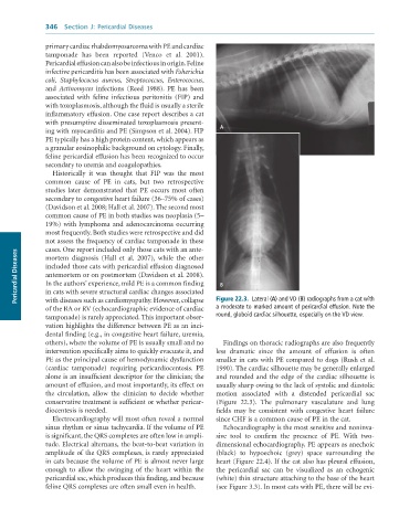
                                                                 a	moderate	to	marked	amount	of	pericardial	effusion.	Note	the
              of the RA or RV (echocardiographic evidence of cardiac   Figure 22.3.  Lateral	(A)	and	VD	(B)	radiographs	from	a	cat	with
              tamponade) is rarely appreciated. This important obser-  round,	globoid	cardiac	silhouette,	especially	on	the	VD	view.
              vation highlights the difference between PE as an inci-
              dental finding (e.g., in congestive heart failure, uremia,
              others), where the volume of PE is usually small and no   Findings on thoracic radiographs are also frequently
              intervention specifically aims to quickly evacuate it, and   less  dramatic  since  the  amount  of  effusion  is  often
              PE as the principal cause of hemodynamic dysfunction   smaller in cats with PE compared to dogs (Rush et al.
              (cardiac  tamponade)  requiring  pericardiocentesis.  PE   1990). The cardiac silhouette may be generally enlarged
              alone is an insufficient descriptor for the clinician; the   and rounded and the edge of the cardiac silhouette is
              amount of effusion, and most importantly, its effect on   usually sharp owing to the lack of systolic and diastolic
              the  circulation,  allow  the  clinician  to  decide  whether   motion  associated  with  a  distended  pericardial  sac
              conservative treatment is sufficient or whether pericar-  (Figure  22.3).  The  pulmonary  vasculature  and  lung
              diocentesis is needed.                             fields  may  be  consistent  with  congestive  heart  failure
                 Electrocardiography will most often reveal a normal   since CHF is a common cause of PE in the cat.
              sinus rhythm or sinus tachycardia. If the volume of PE   Echocardiography is the most sensitive and noninva-
              is significant, the QRS complexes are often low in ampli-  sive  tool  to  confirm  the  presence  of  PE.  With  two-
              tude. Electrical alternans, the beat-to-beat variation in   dimensional echocardiography, PE appears as anechoic
              amplitude of the QRS complexes, is rarely appreciated   (black)  to  hypoechoic  (grey)  space  surrounding  the
              in cats because the volume of PE is almost never large   heart (Figure 22.4). If the cat also has pleural effusion,
              enough to allow the swinging of the heart within the   the  pericardial  sac  can  be  visualized  as  an  echogenic
              pericardial sac, which produces this finding, and because   (white) thin structure attaching to the base of the heart
              feline QRS complexes are often small even in health.  (see Figure 3.3). In most cats with PE, there will be evi-

