Page 334 - Feline Cardiology
P. 334
Chapter 22: Pericardial Effusion and Other Disorders  345


                                                                 initiated  in  a  physiologic  attempt  to  maintain  cardiac
                                                                 output.  Thus,  sinus  tachycardia,  sodium  and  water
                                                                 retention,  and  venoconstriction  help  compensate  for
                                                                 decreased cardiac output. However, increased systemic
                                                                 venous  pressure  leads  to  fluid  transudation  from  sys-
                                                                 temic  capillaries.  Ascites  accumulates  once  central
                                                                 venous  pressure  increases  to  ≥15 mmHg  (normal
                                                                 ≤6 mmHg).  Signs  of  right  heart  failure  predominate
                                                                 because the right ventricle is thinner-walled and more
                                                                 influenced  by  high  intrapericardial  pressures.  Pleural
                                                                 fluid  accumulates  secondary  to  venous  hypertension
                                                                 (similar to ascites), and reduced absorption of subpleu-
                                                                 ral pulmonary lymphatic vessels. However, pulmonary
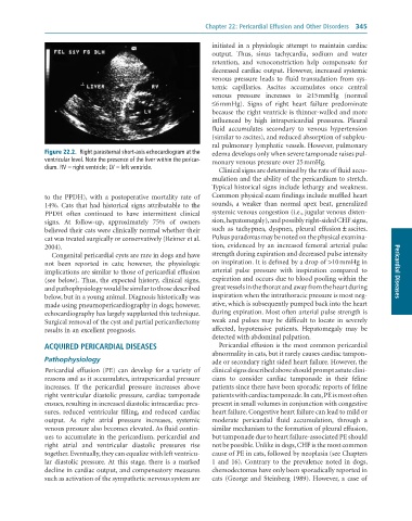
              Figure 22.2.  Right	parasternal	short-axis	echocardiogram	at	the	  edema develops only when severe tamponade raises pul-
              ventricular	level.	Note	the	presence	of	the	liver	within	the	pericar-  monary venous pressure over 25 mmHg.
              dium.	RV	=	right	ventricle;	LV	=	left	ventricle.
                                                                   Clinical signs are determined by the rate of fluid accu-
                                                                 mulation and the ability of the pericardium to stretch.
                                                                 Typical historical signs include lethargy and weakness.
              to  the  PPDH),  with  a  postoperative  mortality  rate  of   Common physical exam findings include muffled heart
              14%. Cats that had historical signs attributable to the   sounds,  a  weaker  than  normal  apex  beat,  generalized
              PPDH  often  continued  to  have  intermittent  clinical   systemic venous congestion (i.e., jugular venous disten-
              signs.  At  follow-up,  approximately  75%  of  owners   sion, hepatomegaly), and possibly right-sided CHF signs,
              believed their cats were clinically normal whether their   such  as  tachypnea,  dyspnea,  pleural  effusion ± ascites.
              cat was treated surgically or conservatively (Reimer et al.   Pulsus paradoxus may be noted on the physical examina-
              2004).                                             tion,  evidenced  by  an  increased  femoral  arterial  pulse
                 Congenital pericardial cysts are rare in dogs and have   strength during expiration and decreased pulse intensity
              not  been  reported  in  cats;  however,  the  physiologic   on inspiration. It is defined by a drop of >10 mmHg in
              implications are similar to those of pericardial effusion   arterial  pulse  pressure  with  inspiration  compared  to   Pericardial Diseases
              (see  below).  Thus,  the  expected  history,  clinical  signs,   expiration and occurs due to blood pooling within the
              and pathophysiology would be similar to those described   great vessels in the thorax and away from the heart during
              below, but in a young animal. Diagnosis historically was   inspiration when the intrathoracic pressure is most neg-
              made using pneumopericardiography in dogs; however,   ative, which is subsequently pumped back into the heart
              echocardiography has largely supplanted this technique.   during expiration. Most often arterial pulse strength is
              Surgical removal of the cyst and partial pericardiectomy   weak  and  pulses  may  be  difficult  to  locate  in  severely
              results in an excellent prognosis.                 affected,  hypotensive  patients.  Hepatomegaly  may  be
                                                                 detected with abdominal palpation.
              ACQUIRED PERICARDIAL DISEASES                        Pericardial effusion is the most common pericardial
                                                                 abnormality in cats, but it rarely causes cardiac tampon-
              Pathophysiology                                    ade or secondary right sided heart failure. However, the
              Pericardial  effusion  (PE)  can  develop  for  a  variety  of   clinical signs described above should prompt astute clini-
              reasons and as it accumulates, intrapericardial pressure   cians  to  consider  cardiac  tamponade  in  their  feline
              increases.  If  the  pericardial  pressure  increases  above   patients since there have been sporadic reports of feline
              right ventricular diastolic pressure, cardiac tamponade   patients with cardiac tamponade. In cats, PE is most often
              ensues, resulting in increased diastolic intracardiac pres-  present in small volumes in conjunction with congestive
              sures,  reduced  ventricular  filling,  and  reduced  cardiac   heart failure. Congestive heart failure can lead to mild or
              output.  As  right  atrial  pressure  increases,  systemic   moderate  pericardial  fluid  accumulation,  through  a
              venous pressure also becomes elevated. As fluid contin-  similar mechanism to the formation of pleural effusion,
              ues to accumulate in the pericardium, pericardial and   but tamponade due to heart failure-associated PE should
              right  atrial  and  ventricular  diastolic  pressures  rise   not be possible. Unlike in dogs, CHF is the most common
              together. Eventually, they can equalize with left ventricu-  cause of PE in cats, followed by neoplasia (see Chapters
              lar  diastolic  pressure.  At  this  stage,  there  is  a  marked   1  and  16).  Contrary  to  the  prevalence  noted  in  dogs,
              decline in cardiac output, and compensatory measures   chemodectomas have only been sporadically reported in
              such as activation of the sympathetic nervous system are   cats  (George  and  Steinberg  1989).  However,  a  case  of

