Page 337 - Feline Cardiology
P. 337
348  Section J: Pericardial Diseases


                Box 22.1.  Procedure	for	pericardiocentesis	in	cats

                1.	 Approach	from	the	left	or	right	side—each	direction	has	  3.	 Perform	a	sterile	prep	of	the	skin.
                  advantages	and	drawbacks;	in	the	example	here,	the	cat	  Infiltrate	the	skin,	intercostal	muscles,	and	parietal
                  is	in	right	lateral	recumbency,	allowing	an	approach	to	the	  pleura	with	local	anesthetic	(2%	lidocaine,	1	ml)	and
                  pericardium	from	the	left	side	unless	echocardiography	  repeat	surgical	preparation.
                  suggests	the	fluid	pocket	is	larger	on	the	right	side.	Mild	  4.	 Use	an	18–20	gauge	over-the-needle	catheter	system	or
                  sedation	can	be	achieved	with	0.3	mg/kg	butorphanol	  a	19	gauge	butterfly	needle	in	a	cat.	Make	two	small	side
                  and	0.3	mg/kg	midazolam	IV	or	IM,	if	necessary.	An	  holes	near	the	tip	of	the	catheter	to	avoid	blockages	during
                  electrocardiogram	should	be	monitored	during	the	  aspiration.
                  procedure	to	monitor	for	ventricular	ectopy	(which	can	  5.	 Attach	the	catheter	to	a	12–20	cc	syringe	via	an	extension
                  occur	if	the	epicardium	is	abraded).             tube	and	a	three-way	stopcock.	If	a	butterfly	needle	is
                    Note:	The	direction	of	approach	for	pericardiocentesis	  used,	you	can	attach	it	directly	to	a	three-way	stopcock	and
                  is	primarily	driven	by	location	of	the	largest	fluid	pocket	  syringe.
                  and	personal	experience.	Some	clinicians	prefer	to	perform	  6.	 Slowly	advance	the	catheter	or	butterfly	needle	through
                  the	procedure	from	the	right	believing	there	is	less	risk	of	  the	skin	toward	the	pericardium	while	maintaining	slight
                  lacerating	a	coronary	artery.	Others	prefer	to	approach	from	  negative	pressure	via	the	syringe.	Once	a	flashback	of
                  the	left	because	deoxygenated	blood	in	the	right	ventricle	  fluid	is	noted,	advance	the	catheter	over	the	needle	into
                  can	easily	be	mistaken	for	the	“port	wine”	appearance	of	  the	pericardial	space	and	remove	the	needle	while	the
                  the	classic	hemorrhagic	pericardial	effusion.	Therefore,	  extension	tube	is	connected	to	the	catheter	(or	advance
                  when	performing	the	procedure	from	the	left,	the	clinician	  the	butterfly	cautiously	toward	the	opposite	scapula).	It
                  knows	quickly	whether	the	sample	is	blood	from	the	left	  is	important	to	keep	in	mind	that	an	initial	flashback	will
                  ventricle	(oxygenated	and	bright	red	in	color),	or	pericardial	  be	pleural	effusion	if	the	patient	has	both	pleural	and
                  fluid	(“port	wine”	(dark	red)	in	color).	As	long	as	the	  pericardial	effusions	present.	Therefore,	the	needle/catheter
                  pericardiocentesis	is	performed	from	the	apex	of	the	heart	  will	need	to	be	advanced	further	for	a	second	flashback,
                                                                   revealing	fluid	that	often	is	different	in	appearance.
                  and	the	needle	is	directed	toward	the	opposite	shoulder,	  7.	 Pericardial	fluid	is	often	less	sanguineous	in	cats
      Pericardial Diseases  2.	 Shave	the	left	(or	right)	thorax	after	the	ideal	location	  compared	to	dogs,	but	if	the	appearance	is	bloody,	place
                  the	risk	of	coronary	laceration	is	low,	whether	one	performs
                  the	procedure	from	the	right	or	left	thorax.
                                                                   an	aliquot	of	fluid	in	an	activated	clotting	time	tube	to
                                                                   confirm	that	it	is	not	blood.	Pericardial	effusion	should	not
                  for	the	procedure	is	determined	by	echocardiography.	In
                                                                   clot,	whereas	blood	from	a	great	vessel	or	cardiac	puncture
                  many	cats,	the	fluid	pocket	is	small	and	echocardiographic
                                                                   will	clot.
                  guidance	during	the	procedure	is	helpful.


                                                                 equilibration of atrial and ventricular diastolic pressures
                                                                 and loss of the normal reduction in caval pressure asso-
                                                                 ciated with inspiration. Right-sided heart failure is the
                                                                 most common abnormality noted on physical examina-
                                                                 tion. This disease likely is extremely rare in the cat, and
                                                                 has not yet been reported.

                                                                 REFERENCES

                                                                 Boldface font indicates key references.
                                                                 Brummer DG, Moïse NS. Infiltrative cardiomyopathy responsive to
                                                                   combination  chemotherapy  in  a  cat  with  lymphoma.  J Am Vet
                                                                   Med Assoc 1989;195:1116–1119.
                                                                 Davidson BJ, Paling AC, Lahmers SL, Nelson OL. Disease associa-
                                                                   tion and clinical assessment of feline pericardial effusion. J Am
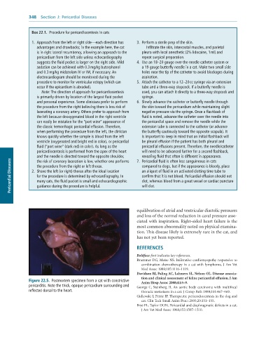
              Figure 22.5.  Postmortem	specimen	from	a	cat	with	constrictive	  Anim Hosp Assoc 2008;44:5–9.
              pericarditis.	Note	the	thick,	opaque	pericardium	surrounding	and	  George  C,  Steinberg  H. An  aortic  body carcinoma  with  multifocal
              reflected	dorsal	to	the	heart.                       thoracic metastases in a cat. J Comp Path 1989;101:467–469.
                                                                 Gidlewski J, Petrie JP. Therapeutic pericardiocentesis in the dog and
                                                                   cat. Clin Tech Small Anim Pract 2005;20:151–155.
                                                                 Frye FL, Taylor DON. Pericardial and diaphragmatic defects in a cat.
                                                                   J Am Vet Med Assoc 1968;152:1507–1510.

