
Page 133 - Basic Monitoring in Canine and Feline Emergency Patients
P. 133
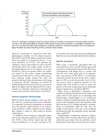
ETCO 2
                       (mmHg)               The alveolar plateau is blunted and blends
  VetBooks.ir              60
                                            with the downstroke of the capnogram
                           50
                           40
                           30
                           20
                           10
                            0
                                                        Time
             Fig. 6.13.  Cardiogenic oscillations. Note the irregular shape of the phase III and phase 0. The wavy shape of phase
             0 is due to the heart transmitting its vibrations to the lung and airway during inspiration. If cardiogenic oscillations are
             seen in an anesthetized patient with spontaneous breathing, assisted or mechanical ventilation may be considered to
             adjust the depth and rate of breathing. ETCO , end-tidal carbon dioxide.
                                             2

             always be interpreted in conjunction with other   circuit and to the water trap on the capnograph and
             physiological variables and the clinical situation.   that the water trap is firmly attached to the machine.
             The ETCO  does NOT replace arterial and venous
                      2
             blood gas analysis.  As mentioned above, at con-
             stant ventilation the ETCO  will represent the   Specific drawbacks
                                    2
             ventilatory status of the patient as well as provide   When using a  colorimetric capnogram, there are
             information about the cardiac output. If there is   some limitations. First, the pH-sensitive paper will
             lung pathology present or the patient is very sick,   respond to anything that changes its pH. Therefore,
             the ETCO  will not accurately reflect the ventila-  contamination with things such as vomitus will
                     2
             tory status nor the cardiac output, necessitating   alter the color of the paper and not be appropri-
             measurement of the PaCO  as well as other param-  ately representative of the ETCO  A colorimetric
                                                                                    2.
                                 2
             eters assessing cardiac output.  The sicker  the   capnogram cannot be used during chest compres-
             patient is, the more likely the perfusion to the lungs   sions in CPR as it will not provide a level of CO .
                                                                                                2
             is negatively affected.  Therefore, in critically ill   The rescuer needs a numerical quantity of CO
                                                                                                2
             intubated patients, the patient’s PaCO  is at least as   exhaled with each breath in order to assess the
                                          2
             high if not higher than the displayed ETCO  meas-  quality of the chest compressions and to determine
                                               2
             urement. It is therefore important to document the   return of spontaneous circulation (ROSC).
             actual carbon dioxide concentration in the blood.  When using a quantitative capnogram, each
                                                         method of sampling (mainstream versus sidestream)
                                                         comes with its own unique set of advantages and
             General equipment shortcomings
                                                         disadvantages.  Disadvantages  of  the  mainstream
             With all carbon dioxide analyzers, water can inter-  analyzers include increased dead space (the analyzer
             fere with CO  analysis and lead to errors. Therefore,   is part of the breathing circuit) and sometimes mild
                       2
             it is important to prevent condensation of moisture   false elevation in CO  readings secondary to accumu-
                                                                         2
             from  the  patient’s  expired  breath  on  the  analyzer   lation  of  condensation  inside  the  unit.  Also,  the
             (some units have a sensor within the airway adapter   weight of the analyzer probe may cause accidental
             that contains a heater or other means to prevent   disconnection from the anesthesia circuit, especially
             condensation).                              with a small ETT.
               In addition, loose connections (especially impor-  Disadvantages of sidestream analyzers include a
             tant in the sidestream analyzer) can lead to falsely   time delay in the waveform (allowing for the time to
             low ETCO  values.  Always ensure that the side-  suction the gas sample from the anesthesia circuit
                      2
             stream sampling line is securely connected to the   and analyze it; this is done at a fixed rate per minute).

             Capnography                                                                     125