
Page 129 - Basic Monitoring in Canine and Feline Emergency Patients
P. 129
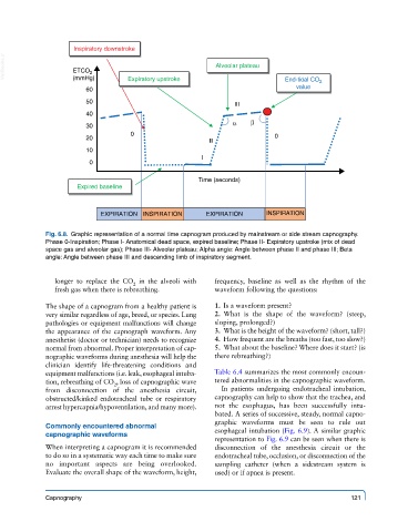
Inspiratory downstroke
  VetBooks.ir       ETCO 2                                Alveolar plateau

                    (mmHg)
                                                                               value
                        60         Expiratory upstroke                      End-tidal CO 2
                        50                                     III
                        40
                        30                                    α    β
                                   0                                     0
                        20
                                                        II
                        10
                                                      I
                         0

                                                     Time (seconds)
                     Expired baseline


                           EXPIRATION  INSPIRATION     EXPIRATION      INSPIRATION


             Fig. 6.8.  Graphic representation of a normal time capnogram produced by mainstream or side stream capnography.
             Phase 0-Inspiration; Phase I- Anatomical dead space, expired baseline; Phase II- Expiratory upstroke (mix of dead
             space gas and alveolar gas); Phase III- Alveolar plateau; Alpha angle: Angle between phase II and phase III; Beta
             angle: Angle between phase III and descending limb of inspiratory segment.

               longer to replace the CO  in the alveoli with   frequency, baseline as well as the rhythm of the
                                    2
               fresh gas when there is rebreathing.      waveform following the questions:

             The shape of a capnogram from a healthy patient is     1.  Is a waveform present?
             very similar regardless of age, breed, or species. Lung     2.  What is the shape of the waveform? (steep,
             pathologies or equipment malfunctions will change     sloping, prolonged?)
             the appearance of the capnograph waveform. Any     3.  What is the height of the waveform? (short, tall?)
             anesthetist (doctor or technician) needs to recognize     4.  How frequent are the breaths (too fast, too slow?)
             normal from abnormal. Proper interpretation of cap-    5.  What about the baseline? Where does it start? (is
             nographic waveforms during anesthesia will help the   there rebreathing?)
             clinician identify life-threatening conditions and
             equipment malfunctions (i.e. leak, esophageal intuba-  Table 6.4 summarizes the most commonly encoun-
             tion, rebreathing of CO , loss of capnographic wave   tered abnormalities in the capnographic waveform.
                               2
             from disconnection of the anesthesia circuit,   In patients undergoing endotracheal intubation,
             obstructed/kinked endotracheal tube or respiratory   capnography can help to show that the trachea, and
             arrest hypercapnia/hypoventilation, and many more).  not the esophagus, has been successfully intu-
                                                         bated. A series of successive, steady, normal capno-
                                                         graphic waveforms must be seen to rule out
             Commonly encountered abnormal
             capnographic waveforms                      esophageal intubation (Fig. 6.9). A similar graphic
                                                         representation to Fig. 6.9 can be seen when there is
             When interpreting a capnogram it is recommended   disconnection of the anesthesia circuit or the
             to do so in a systematic way each time to make sure   endotracheal tube, occlusion, or disconnection of the
             no important aspects are being overlooked.   sampling catheter (when a sidestream system is
             Evaluate the overall shape of the waveform, height,   used) or if apnea is present.


             Capnography                                                                     121