
Page 134 - Basic Monitoring in Canine and Feline Emergency Patients
P. 134
Also, as mentioned previously, sidestream units distorted by the fresh gas flow. However, when
require connection to a scavenging system in order using a catheter or needle, there can be disconnec-
VetBooks.ir to remove inhaled anesthetic gases. As it removes tion at the site of insertion into the ETT or obstruc-
tion of the catheter or needle by secretions or
inhaled anesthetic gas from the patient, there can
uncommonly be inadvertent exposure of the envi-
discarded after use if the catheter or needle has cre-
ronment and the staff to the gas. Finally, the small bending. In rare cases, the ETT may have to be
sampling tubing can become obstructed with water, ated a leak in the tube. A third alternative is to use
blood or secretions and become non-functional. an airway adapter that has an inner lumen with a
In addition, small patients (less than 7 kg) are small diameter (Fig. 6.15). The smaller diameter of
usually connected to a non-rebreathing anesthesia the inner lumen reduces the dead space of the
system which uses a high fresh gas flow. This can adapter as it fits perfectly to the ETT connector
result in distortion of the sidestream capnograph through which the patient’s gas stream will pass.
waveform leading to an erroneously low ETCO This adaptor is easier to use than an additional
2
reading. Several options exist to overcome this needle or catheter and carries a minimal risk of
limitation. One alternative is to use the lowest pos- blockage or contamination by secretions. This type
sible sampling rate on your monitor. A second of airway adaptor is commonly used in exotics.
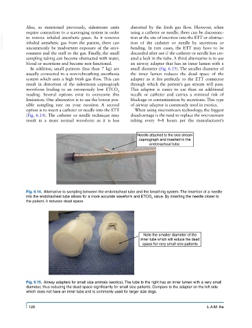
option is to insert a catheter or needle into the ETT When using microstream technology, the biggest
(Fig. 6.14). The catheter or needle technique may disadvantage is the need to replace the microstream
result in a more normal waveform as it is less tubing every 4–8 hours per the manufacturer’s
Needle attached to the side stream
capnograph and inserted in the
endotracheal tube.
Fig. 6.14. Alternative to sampling between the endotracheal tube and the breathing system. The insertion of a needle
into the endotracheal tube allows for a more accurate waveform and ETCO value. By inserting the needle closer to
2
the patient, it reduces dead space.
Note the smaller diameter of the
inner tube which will reduce the dead
space for very small size patients
Fig. 6.15. Airway adapters for small size animals (exotics). The tube to the right has an inner lumen with a very small
diameter, thus reducing the dead space significantly for small size patients. Compare to the adapter on the left side
which does not have an inner tube and is commonly used for larger size dogs.
126 L.A.M. Ilie