
Page 130 - Basic Monitoring in Canine and Feline Emergency Patients
P. 130
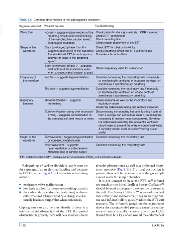
Table. 6.4.  Common abnormalities in the capnographic waveform.

  VetBooks.ir  Segment affected  Possible causes        Troubleshooting
             Wave form
                           Absent – suggests disconnection of the  Check patient’s vital signs and start CPR if needed
                             breathing circuit; obstruction/kinking
                                                         Check sampling line
                             of the sampling line; cardiac arrest;   Check ETT connections
                             esophageal intubation       Check proper placement of the ETT
             Shape of the   Slant (prolonged) phase II or III –   Check ETT for obstruction/kinks
              waveform       suggests obstruction of the expiratory  Check breathing circuit and ETT cuff for leaks
                             flow (i.e kinked ETT; bronchospasm;   Consider a bronchodilator
                             asthma) or leaks in the breathing
                             system
                           Slant (prolonged) phase 0 – suggests
                             malfunction of the inspiratory valve   Check inspiratory valve for malfunction
                             when a closed-circuit system is used
             Frequency of    Too fast – suggests hyperventilation  Consider decreasing the respiratory rate if manually
              the waveform                                or mechanically ventilated or increase the depth of
                                                          anesthesia if spontaneously breathing
                           Too slow – suggests hypoventilation  Consider increasing the respiratory rate if manually
                                                          or mechanically ventilated or reduce depth of
                                                          anesthesia if spontaneously breathing
             Inspiratory    Gradual elevation – suggests   Check sodalime as well as the inspiratory and
              baseline       rebreathing                  expiratory valves
                                                         Check the sidestream tubing and replace if needed
                           Sudden elevation along with increased  Disconnecting the sampling line and flushing it with air
                             ETCO  – suggests contamination of   from a syringe can sometimes clear it, but it may be
                                 2
                             the sampling cell with mucus or water  necessary to replace these components. Elevating
                                                          the sidestream sampling line above the ventilator
                                                          circuit helps to prevent the entry of condensed water.
                                                          A humidity barrier such as Nafion  tubing is also
                                                                                  ®
                                                          useful
             Height of the   Tall waveform – suggests hypoventilation  Consider increasing the respiratory rate
              waveform       or increased metabolic rate
                           Short waveform – suggests     Consider decreasing the respiratory rate
                             hyperventilation or a decrease in
                             metabolic rate or cardiac output
             ETT, endotracheal tube; CPR, cardiopulmonary resuscitation; ETCO , end-tidal carbon dioxide.
                                                       2
              Rebreathing of carbon dioxide is easily seen on   alveolar plateau noted as well as a prolonged inspir-
            the capnogram as an elevated baseline and increase   atory upstroke (Fig. 6.11). If a total obstruction is
            in ETCO  value (Fig. 6.10). Causes for rebreathing   present, there will be no waveform as the gas sample
                   2
            include:                                     cannot reach the sample chamber.
                                                          It is not unusual to have the ETT cuff inflated
             ● ● expiratory valve malfunction;           too much or too little. Ideally a Possey Cufflator TM
             ● ● low fresh gas flow (in the non-rebreathing  circuits);   should be used to properly measure the pressure in
             ● ● the carbon dioxide absorber (soda lime) is chemi-  the cuff. The Possey Cufflator TM  is an endotracheal
               cally exhausted (characterized by a change in color -    tube inflator and manometer. It has an air vent but-
               usually becomes purple/blue when exhausted).  ton and inflator bulb to quickly adjust the ETT cuff
                                                         pressure.  The inflator’s gauge on the manometer
            Capnograms can also help us identify if there is a   shows the recommended pressure range in centim-
            total or partial obstruction of the ETT. If a partial   eters of water (usually between 20–30  cm H O).
                                                                                             2
            obstruction is present, there will be a small or absent   Should there be a leak of air around the endotracheal

             122                                                                         L.A.M. Ilie