
Page 741 - Small Animal Internal Medicine, 6th Edition
P. 741
CHAPTER 43 Canine and Feline Urolithiasis 713
often not as rewarding but can be attempted. It is important bladder expressions. A urinary catheter is then inserted
when attempting to dissolve struvite stones in dogs, in addi- into the bladder, and the bladder should be distended with
VetBooks.ir tion to dietary therapy (see later), to use appropriate antimi- sterile saline. The bladder should feel full but not so taut
that bladder rupture could occur. With the catheter still in
crobial therapy as well.
VUH can be performed when small calculi are present
bral column is vertical, and the bladder should be agitated
in the bladder (Fig. 43.5). The urolith size and shape, as place, the animal should be held upright so that the verte-
well as the size of the dog or cat, need to be assessed; to help promote movement of the stones into the bladder
smoother stones generally pass through the smallest diam- neck. As soon as the catheter is removed, the clinician should
eter of the urethra easier than those with irregular surfaces. express the bladder to create a forceful stream and collect the
Large uroliths can be removed more easily from larger voided contents. When expressing the bladder, the palm of
dogs, although the procedure can be more cumbersome. To the hand (not the fingertips) should be used to help prevent
perform a VUH, the dog or cat should be anesthetized to trauma to the bladder. Several voids may be required to
help prevent increased urethral pressure and facilitate easier remove all the stones and stone debris present. Hematuria
+
+
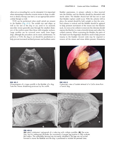
FIG 43.1 FIG 43.3
Ultrasonogram of a large cystolith in the bladder of a dog. Cystoscopic view of basket retrieval of a CaOx stone from
Note the intense shadowing produced by the urolith. a female dog.
A B
FIG 43.2
(A) Lateral abdominal radiograph of a male dog with multiple cystoliths. (B) The same
dog, but this radiograph illustrates the importance of proper leg position to fully evaluate
the urethra. The urethroliths (arrows) are not easily identified if the legs are not pulled
forward to move the fabellae and the dog is positioned to include the perineal area.