
Page 743 - Small Animal Internal Medicine, 6th Edition
P. 743
CHAPTER 43 Canine and Feline Urolithiasis 715
diagnostic imaging in cats with renal disease. Ureterolithiasis
tends to develop in middle-aged to older cats, with a median
VetBooks.ir age of 7 years at the time of diagnosis.
Although reported less often, other causes of ureteral
obstruction can include soft tissue plugs, which sometimes
contain flakes of mineralized material, inflammatory debris
in cats with pyelonephritis, and calculi composed of 100%
dried solidified blood (DSB). In cats with a chronic upper
tract urolithiasis, in which calculi have passed previously, it
is relatively common for significant ureteral inflammation
and/or stricture to develop, further decreasing the luminal
diameter through which material can pass. This can lead to
an even higher risk for the development of ureteral obstruc-
tion with minerals, whereby debris that would pass in a
normal ureter leads to obstruction.
CLINICAL SIGNS OF URETEROLITHIASIS
Because ureterolithiasis is much more common in cats com-
pared with dogs, the discussion will focus on this species,
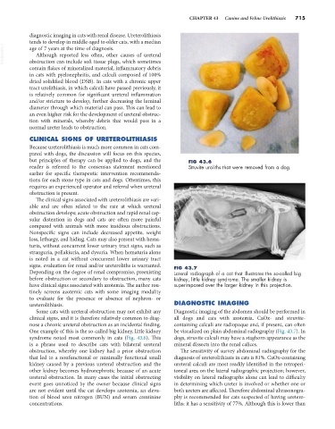
but principles of therapy can be applied to dogs, and the FIG 43.6
reader is referred to the consensus statement mentioned Struvite uroliths that were removed from a dog.
earlier for specific therapeutic intervention recommenda-
tions for each stone type in cats and dogs. Oftentimes, this
requires an experienced operator and referral when ureteral
obstruction is present.
The clinical signs associated with ureterolithiasis are vari-
able and are often related to the rate at which ureteral
obstruction develops; acute obstruction and rapid renal cap-
sular distention in dogs and cats are often more painful
compared with animals with more insidious obstructions.
Nonspecific signs can include decreased appetite, weight
loss, lethargy, and hiding. Cats may also present with hema-
turia, without concurrent lower urinary tract signs, such as
stranguria, pollakiuria, and dysuria. When hematuria alone
is noted in a cat without concurrent lower urinary tract
signs, evaluation for renal and/or ureteroliths is warranted. FIG 43.7
Depending on the degree of renal compromise, preexisting Lateral radiograph of a cat that illustrates the so-called big
before obstruction or secondary to obstruction, many cats kidney, little kidney syndrome. The smaller kidney is
have clinical signs associated with azotemia. The author rou- superimposed over the larger kidney in this projection.
tinely screens azotemic cats with some imaging modality
to evaluate for the presence or absence of nephron- or
ureterolithiasis. DIAGNOSTIC IMAGING
Some cats with ureteral obstruction may not exhibit any Diagnostic imaging of the abdomen should be performed in
clinical signs, and it is therefore relatively common to diag- all dogs and cats with azotemia. CaOx- and struvite-
nose a chronic ureteral obstruction as an incidental finding. containing calculi are radiopaque and, if present, can often
One example of this is the so-called big kidney, little kidney be visualized on plain abdominal radiography (Fig. 43.7). In
syndrome noted most commonly in cats (Fig. 43.6). This dogs, struvite calculi may have a staghorn appearance as the
is a phrase used to describe cats with bilateral ureteral mineral dissects into the renal calices.
obstruction, whereby one kidney had a prior obstruction The sensitivity of survey abdominal radiography for the
that led to a nonfunctional or minimally functional small diagnosis of ureterolithiasis in cats is 81%. CaOx-containing
kidney caused by a previous ureteral obstruction and the ureteral calculi are most readily identified in the retroperi-
other kidney becomes hydronephrotic because of an acute toneal area on the lateral radiographic projection; however,
ureteral obstruction. In many cases the initial obstructing visibility on lateral radiographs alone can lead to difficulty
event goes unnoticed by the owner because clinical signs in determining which ureter is involved or whether one or
are not evident until the cat develops azotemia, an eleva- both ureters are affected. Therefore abdominal ultrasonogra-
tion of blood urea nitrogen (BUN) and serum creatinine phy is recommended for cats suspected of having uretero-
concentrations. liths; it has a sensitivity of 77%. Although this is lower than