
Page 745 - Small Animal Internal Medicine, 6th Edition
P. 745
CHAPTER 43 Canine and Feline Urolithiasis 717
BOX 43.1
VetBooks.ir Calcium Oxalate Stone Management for Lower
Urinary Urolithiasis
1. Remove all uroliths; obtain radiographs to be certain
all uroliths have been removed.
2. Submit all uroliths for quantitative crystallographic
analysis.
3. Evaluate a serum calcium concentration.
• If high, pursue further diagnostics, such as an
ionized calcium and parathyroid hormone (PTH)
panel.
4. Consider evaluating serum triglyceride concentrations.
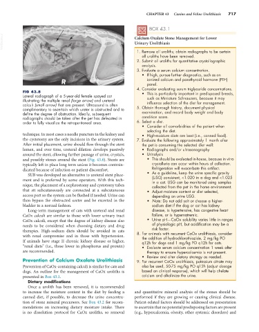
FIG 43.8 • This is particularly important in predisposed breeds,
Lateral radiograph of a 5-year-old female spayed cat such as Miniature Schnauzers, because it may
illustrating the multiple renal (large arrow) and ureteral
calculi (small arrow) that are present. Ultrasound is often influence selection of the diet for management.
complimentary to ascertain which ureter is obstructed and to 5. Obtain thorough history, document physical
define the degree of obstruction. Ideally, subsequent examination, and record body weight and body
radiographs should be taken after the pet has defecated in condition score.
order to fully visualize the retroperitoneal area. 6. Select a diet.
• Consider all comorbidities of the patient when
selecting the diet.
technique. In most cases a needle puncture in the kidney and • High-moisture diets are best (i.e., canned food).
the cystotomy are the only incisions in the urinary system. 7. Evaluate the following approximately 1 month after
After initial placement, urine should flow through the stent the pet is consuming the selected diet well:
lumen, and over time, ureteral dilation develops passively • Radiographs and/or ultrasonography
around the stent, allowing further passage of urine, crystals, • Urinalysis
and possibly stones around the stent (Fig. 43.8). Stents are • This should be evaluated in-house, because in vitro
typically left in place long term unless it becomes contrain- crystalluria can occur within hours of collection.
dicated because of infection or patient discomfort. Refrigeration will exacerbate this artifact.
SUB was developed an alternative to ureteral stent place- • As a guideline, keep the urine specific gravity
ment and is preferred by some operators. With this tech- (USG) consistent, <1.020 in a dog and <1.025
in a cat. USG can be monitored using samples
nique, the placement of a nephrostomy and cystotomy tubes collected from the pet in the home environment.
that sit subcutaneously are connected at a subcutaneous • Adjust moisture content or diet selected,
access port so the system can be flushed if needed. Urine can depending on urine USG.
then bypass the obstructed ureter and be excreted in the • Note: Do not add salt or choose a higher-
bladder in a normal fashion. sodium diet if the dog or cat has kidney
Long-term management of cats with ureteral and renal disease, is hypertensive, has congestive heart
CaOx calculi are similar to those with lower urinary tract failure, or is hypernatremic.
CaOx calculi, except that the degree of kidney disease also • Urine pH—CaOx solubility varies little in ranges
needs to be considered when choosing dietary and drug of physiologic pH, but acidification may be a
risk factor.
therapies. High-sodium diets should be avoided in cats 8. For animals with recurrent CaOx urolithiasis, consider
with renal compromise and in those with hypertension. the addition of hydrochlorothiazide, 2 mg/kg PO
If animals have stage II chronic kidney disease or higher, q12h for dogs and 1 mg/kg PO q12h for cats.
“renal diets” (i.e., those lower in phosphorus and protein) • Evaluate serum calcium concentration 1 week after
are recommended. therapy to ensure hypercalcemia is not present.
• Review and alter dietary strategy as needed.
Prevention of Calcium Oxalate Urolithiasis 9. For recurrent CaOx urolithiasis, potassium citrate may
Prevention of CaOx-containing calculi is similar for cats and also be used, 50-75 mg/kg PO q12h (adjust dosage
dogs. An outline for the management of CaOx uroliths is based on clinical response), which will help chelate
presented in Box 43.1. calcium and alkalinize the urine.
Dietary modifications
Once a urolith has been removed, it is recommended
to increase the moisture content in the diet by feeding a and quantitative mineral analysis of the stones should be
canned diet, if possible, to decrease the urine concentra- performed if they are growing or causing clinical disease.
tion of stone mineral precursors. See Box 43.2 for recom- Patient-related factors should be addressed on presentation
mendations on increasing dietary moisture intake. There to ascertain that no potential predisposing factors are present
is no dissolution protocol for CaOx uroliths, so removal (e.g., hypercalcemia, obesity, other systemic disorders) and