
Page 742 - Small Animal Internal Medicine, 6th Edition
P. 742
714 PART V Urinary Tract Disorders
CALCIUM OXALATE CALCULI
VetBooks.ir Etiology
CaOx is the most common urolith from dogs submitted to
several veterinary stone laboratories. They appear to be more
common in older, castrated male dogs; small-breed dogs
such as the Bichon Frise, Miniature Schnauzer, Pomeranian,
Cairn terrier, and Maltese are at higher risk for CaOx urolith
formation. The Keeshond has also been reported to have a
higher risk for forming CaOx uroliths, which may be linked
to its genetic predisposition for primary hyperparathyroid-
ism causing hypercalcemia and hypercalciuria. Dogs with
CaOx have been reported to have a higher body condition
score than dogs without CaOx in a recent study.
CaOx is also the most common urolith removed cats. The
most common location for CaOx in both species is the
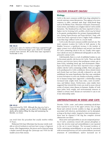
FIG 43.4 bladder, however, a significant increase in the number of
Cystoscopic view of a holmium:YAG laser inserted through upper urinary tract calculi (kidneys and ureters) was found
the scope to fractionate larger cystic calculi for minimally for CaOx-containing calculi in the cat. Usually when upper
invasive stone removal. All uroliths have been reported to
fractionate in vitro. tract calculi are seen on abdominal radiographs in cats, they
primarily contain CaOx.
Unfortunately, there is a lack of published studies in cats
to document specific risk factors for CaOx. There are likely
intrinsic and extrinsic factors that predispose certain cats to
form CaOx, including breed, age, and environment. Hima-
layan and Persian cats have been reported to have a higher
relative risk for CaOx-containing uroliths. Male cats appear
to be predisposed to CaOx urolithiasis. Stress-related events
and obesity have not been critically evaluated in cats with
urolithiasis, but some hypothesize that they may contribute
to stone formation in some cats. Studies evaluating urolithia-
sis in human beings have supported a relationship among
increased body weight, body mass index, waist circumfer-
ence, and kidney stones. Furthermore, some studies have
suggested that stressful life events can result in more clinical
events of urinary stone disease in humans. Studies of body
mass index, body weight, and environmental stressors as
related to urinary stone disease are warranted in cats to see
if any relationship exists.
URETEROLITHIASIS IN DOGS AND CATS
Struvite-, CaOx-, urate-, and cystine-containing calculi have
FIG 43.5
Dog positioned for VUH. Although this dog also had a been reported to occur in the upper urinary tract of dogs.
cystoscopy, a catheter can be used to fill the bladder with Renal and ureteral struvite uroliths in dogs are generally
sterile saline before lifting the dog and expressing the associated with infection, and dissolution can be attempted,
bladder, as shown. as long as the dog is stable. Surgical or interventional proce-
dures may be required if the dog has complete ureteral
obstruction (see later).
can result from this procedure but usually resolves within Most ureter and nephroliths in cats are composed primar-
24 hours. ily of CaOx, sometimes mixed with calcium phosphate or
Holmium:YAG laser lithotripsy has become widely avail- urate, but struvite renal and ureterolithiasis is not reported
able at referral institutions and uses laser energy to fragment in the cat in recent literature. The increased incidence of
stones. After fragmentation, the pieces can be removed using CaOx-containing ureterolithiasis in cats may be associated
a basket inserted through the cystoscope and VUH for the with increased prevalence of CaOx ureterolithiasis, increased
smaller pieces that remain. awareness of ureterolithiasis, and/or increased use of