
Page 390 - Withrow and MacEwen's Small Animal Clinical Oncology, 6th Edition
P. 390
368 PART IV Specific Malignancies in the Small Animal Patient
and histologic parameters. On divergent ends of the malignancy Size and Stage
spectrum, a low-grade 0.5-cm haired-skin melanocytoma is highly For dogs with oral melanoma, primary tumor size has been found
likely to be cured with simple surgical excision whereas a 5.0-cm
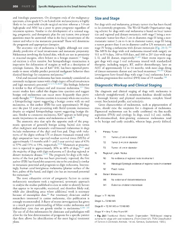
VetBooks.ir high-grade oral MM has a poor to grave prognosis regardless of to be extremely prognostic. The World Health Organization stag-
ing scheme for dogs with oral melanoma is based on local tumor
treatment options. Similar to the development of a rational stag-
ing, prognostic, and therapeutic plan for any tumor, two primary size and regional and distant metastasis, with stage I being a non-
questions must be answered: what is the local invasiveness of the metastatic tumor less than 2 cm in diameter, stage II being a non-
tumor and what is the metastatic potential? These will determine metastatic tumor 2 cm to 4 cm in diameter tumor, stage III being
the prognosis and appropriate therapies. a tumor greater than 4 cm in diameter and/or LN metastasis, and
The anatomic site of melanoma is highly, although not com- stage IV being a melanoma with distant metastasis (Fig. 20.1). 104
pletely, predictive of local invasiveness and metastatic propensity. The MSTs for dogs with oral melanoma treated with surgery are
Melanomas involving the haired skin, which are not in proximity 511 to 874 days, 160 to 818 days, and 168 to 207 days with stage
1,3
to mucosal margins, often behave in a benign manner. Surgi- I, II, and III disease, respectively. 17,105 More recent reports sug-
cal excision is often curative, but histopathologic examination is gest dogs with stage I oral melanoma treated with standardized
imperative for delineation of margins as well as a description of therapies, including surgery, RT, and/or chemotherapy, have an
cytologic features. The use of Ki67 IHC has been reported previ- MST of approximately 12 to 14 months, with most dogs dying
ously to more reliably predict potential malignant behavior than of distant metastatic disease and not local recurrence. 106,107 Other
classical histology for cutaneous melanoma. 89 investigators have found dogs with stage I oral melanoma have a
Oral and mucosal melanoma has been routinely considered an median progression-free survival (PFS) time of 19 months. 108
extremely malignant tumor with a high degree of local invasiveness
and high metastatic potential. 2,13,17,18,30,90 This biologic behavior Diagnostic Workup and Clinical Staging
is similar to that of human oral and mucosal melanoma. 3,91 Two
recent studies have called this dogma into question and suggest The diagnosis and clinical staging of dogs with melanoma is
benign oral melanomas can occur more frequently than previ- relatively straightforward. A minimum database should include
ously thought. 92,93 Extreme caution is necessary when one receives a thorough history and physical examination, complete blood
a histopathology report suggesting a benign course with an oral count, biochemical profile, and urinalysis.
melanoma, as this author (PJB) has seen approximately 30 dogs Gross characteristics of melanomas, such as pigmentation of
over the past 12 years presenting with florid systemic metastases mass, should raise the suspicion for a diagnosis of melanoma.
despite an original histopathology report suggesting benign dis- Pigmented melanomas can be easily confirmed via fine-needle
ease. Similar to cutaneous melanoma, Ki67 appears to hold prog- aspiration (FNA) and cytology. In dogs, small (<2 cm), mobile,
nostic importance in canine oral melanoma as well. 94 well-circumscribed, slow-growing cutaneous melanomas tend
The anatomic sites that split the opposite ends of the prog- to be benign and easily excisable, whereas large, poorly defined,
nostic spectrum of generally benign acting, haired skin versus
often malignant and metastatic oral/mucosal melanomas in dogs
include melanomas of the digit and foot pad. Dogs with mela- T: Primary Tumor
noma of the digits without LN or distant metastasis treated with
digit amputation have reported median survival times (MSTs) of T1 Tumor <2 cm in diameter
approximately 12 months with 1- and 2-year survival rates of 42% T2 Tumor 2–4 cm in diameter
to 57% and 11% to 13%, respectively. 95,96 Metastasis at presenta-
tion is reported in approximately 30% to 40% of dogs, 95,97 and T3 Tumor >4 cm in diameter
the majority of dogs with digit melanoma will develop regional or
distant metastatic disease. 95–97 The prognosis for dogs with mela- N: Regional Lymph Nodes
noma of the foot pad has not been previously reported; the first N0 No evidence of regional node involvement
author (PJB) has found this anatomic site to be anecdotally similar
in metastatic potential and prognosis to digit melanoma. Interest- N1 Histologic/Cytologic evidence of regional node involvement
ingly, human acral lentiginous melanoma (plantar surface of the N2 Fixed nodes
foot, palms of the hand, and digit) also has an increased potential
for metastasis. 98 M: Distant Metastasis
The most exhaustive review of prognostic factors in canine
melanocytic neoplasms took a regimented, systematic approach M0 No evidence of distant metastasis
to analyze the studies published to date in order to identify factors M1 Evidence of distant metastasis
that appear to be repeatable, statistical, and therefore likely real,
while also identifying areas where additional work is necessary
99
because of incomplete data. For veterinary clinicians and/or Stage I = T1 N0 M0
researchers interested in canine melanoma, this publication is
strongly recommended. A flurry of recent investigations has given Stage II = T2 N0 M0
us a much greater understanding of feline ocular melanoma and
iridiociliary cysts that are grossly similar to melanoma. 6,100–103 Stage III = T2 N1 M0 or T3 N0 M0
The use of all this information by clinicians and pathologists will Stage IV = Any T, Any N and M1
allow for the best determination of prognosis for a specific patient • Fig. 20.1 Traditional World Health Organization TNM-based staging
that then allows for identification of the most logical treatment scheme for dogs with oral melanoma. (From Owen LN. TNM Classification
plans. of Tumors in Domestic Animals. 1st ed. Geneva, Switzerland; 1980.)