
Page 391 - Withrow and MacEwen's Small Animal Clinical Oncology, 6th Edition
P. 391
CHAPTER 20 Melanoma 369
ulcerated, and rapidly growing tumors can make surgical excision histopathologic testing. Selective lymphadenectomy avoids the
challenging. In the latter example, incisional biopsy and IHC can indiscriminate extirpation of multiple LNs and, because it is a
less extensive surgical dissection, reduces the risk of postoperative
be an important part of the diagnostic workup, particularly if the
VetBooks.ir mass is nonpigmented, to allow other tumor types to be ruled out complications. The use of SLN mapping and lymphadenectomy
has been proven to be of diagnostic, prognostic, and clinical bene-
and a diagnosis of melanoma confirmed.
Clinical staging tests routinely include evaluation of the regional fit in human melanoma. 115 Relatively few investigations have been
LNs and assessment of the thoracic cavity with either three-view tho- reported to date for SLN mapping and/or excision for dogs with
racic radiographs or thoracic CT scan. The regional LNs should be malignancies, 116–123 and the authors strongly encourage addi-
assessed whether lymphadenomegaly is present or not. LN metas- tional investigation and clinical adoption in this area.
tasis is present in approximately 70% of dogs with lymphadeno- Although not commonly described, abdominal ultrasonogra-
megaly (Fig. 20.2) but, more importantly, in approximately 40% phy or CT scans should be considered in dogs with melanomas
with normal sized LNs. 109 FNA cytology of the ipsilateral regional arising from the oral cavity, digits, or pads because of the risk of
LNs has been recommended to assess for nodal metastasis; how- metastasis to the abdominal LNs, liver, adrenal glands, and other
ever, the only accessible regional LN is the mandibular LN. This intraabdominal sites. The use of novel staging modalities, such as
may provide misleading information, as the lymphatic drainage of gallium citrate scintigraphy, requires further investigation. 124
the head is complex and metastases to contralateral LNs has been Cross-sectional imaging (i.e., computed tomography [CT]
documented. Furthermore, the mandibular LN can be normal scans or magnetic resonance imaging [MRI]) is critical for surgical
when other LNs, such as the medial retropharyngeal or parotid planning for oral melanoma, especially melanomas involving the
LN, are metastatic. 110,111 Although earlier studies showed a high maxilla and caudal mandible. This allows for assessment of the
concordance of cytology and histopathology in dogs with melano- extent of tumor, invasion into soft tissues, bone and the nasal cav-
mas, 111 a recent study highlighted discordance between cytology ity, and assessment of regional LNs, particularly the nonpalpable
findings and histopathology, with a low correlation between the medial retropharyngeal, parotid, and buccal LNs.
final cytology and histopathology reports in dogs with melano-
cytic neoplasia. 112 As a result, histologic examination of the LN
is recommended either through excision of the major LNs of the Treatment
head and neck or sentinel LN (SLN) mapping. Surgery
Nondiscriminate extirpation of the regional LNs of the head
and neck has been described in dogs with and cats with malig- Surgery continues to be the most effective local treatment modal-
nant oral tumors. 113,114 One approach describes ipsilateral extir- ity for melanoma. There are few objective data available to guide
pation of the parotid, mandibular, and retropharyngeal LNs, but decision making for appropriate surgical margin width for resec-
this requires an extensive dissection and does not investigate the tion of melanomas. Benign cutaneous tumors are typically com-
contralateral LNs. 113 A second approach involves extirpation of pletely excised with 1-cm skin margins (and ideally one fascial
the left and right mandibular and medial retropharyngeal LNs plane for deep margins). Given the invasive nature of MMs, wide
through a single incision, but does not investigate the parotid margins (2–3 cm) are ideal whenever possible; however, for oral
LNs. 114 The limitations of both approaches include more exten- melanomas, wide margins may not possible because of the limited
sive dissections and incomplete testing of the regional LNs, and amount of adjacent normal tissues. In these cases, surgery may
hence there is a risk of postoperative complications and missed need to be combined with RT for adequate local tumor control. In
metastatic LNs. the authors’ experience, 1- to 2-cm margins are usually adequate
SLN mapping allows identification of the direct lymphatic for complete histologic excision of MMs with well-defined bor-
drainage pathway from the tumor to the first draining LN. This ders (Fig. 20.3). Wide margins are usually possible with cutaneous
LN can then be targeted for selective lymphadenectomy and and digit melanomas.
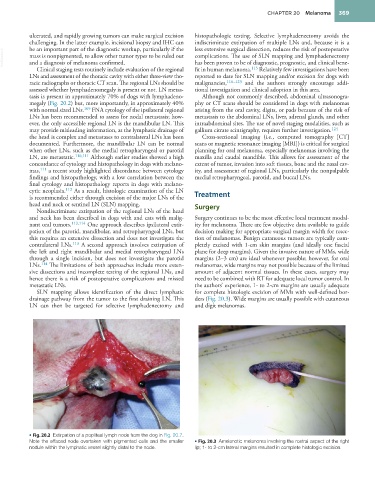
• Fig. 20.2 Extirpation of a popliteal lymph node from the dog in Fig. 20.7.
Note the effaced node overtaken with pigmented cells and the smaller • Fig. 20.3 Amelanotic melanoma involving the rostral aspect of the right
nodule within the lymphatic vessel slightly distal to the node. lip; 1- to 2-cm lateral margins resulted in complete histologic excision.