
Page 392 - Withrow and MacEwen's Small Animal Clinical Oncology, 6th Edition
P. 392
370 PART IV Specific Malignancies in the Small Animal Patient
VetBooks.ir
A
• Fig. 20.4 Local tumor recurrence after incomplete histologic excision
and postoperative radiation of a gingival melanoma dorsal to the right car-
nassial tooth (caudal is to the left and the right canine tooth can be seen in
the lower right-hand corner). When first treated, the mass was reportedly
less than 1 cm in diameter and wide resection (i.e., partial maxillectomy)
was not performed.
Partial mandibulectomy or maxillectomy is often required for
resection of oral melanomas arising from the gingiva or mucosa
in close proximity to the bone. 125 A common error is to resect
a gingival mass without taking underlying bone simply because
bone invasion is not observed. Owing to the proximity of the
gingiva to the underlying bone, this approach typically leaves
residual microscopic disease that leads to local recurrence (Fig.
20.4). Occasionally, tumors do occur in mucosal areas that are not
adjacent to bone (e.g., buccal mucosa) or originate in the lip or B
tongue (Fig. 20.5) and are amenable to excision of the soft tissues
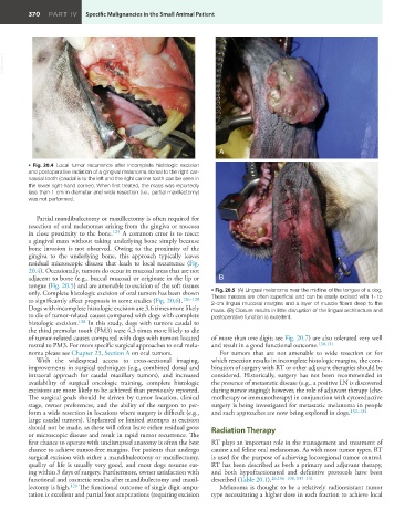
only. Complete histologic excision of oral tumors has been shown • Fig. 20.5 (A) Lingual melanoma near the midline of the tongue of a dog.
to significantly affect prognosis in some studies (Fig. 20.6). 126–128 These masses are often superficial and can be easily excised with 1- to
2-cm lingual mucosal margins and a layer of muscle fibers deep to the
Dogs with incomplete histologic excision are 3.6 times more likely mass. (B) Closure results in little disruption of the lingual architecture and
to die of tumor-related causes compared with dogs with complete postoperative function is excellent.
histologic excision. 128 In this study, dogs with tumors caudal to
the third premolar tooth (PM3) were 4.3 times more likely to die
of tumor-related causes compared with dogs with tumors located of more than one digit; see Fig. 20.7) are also tolerated very well
rostral to PM3. For more specific surgical approaches to oral mela- and result in a good functional outcome. 130,131
noma please see Chapter 23, Section A on oral tumors. For tumors that are not amenable to wide resection or for
With the widespread access to cross-sectional imaging, which resection results in incomplete histologic margins, the com-
improvements in surgical techniques (e.g., combined dorsal and bination of surgery with RT or other adjuvant therapies should be
intraoral approach for caudal maxillary tumors), and increased considered. Historically, surgery has not been recommended in
availability of surgical oncologic training, complete histologic the presence of metastatic disease (e.g., a positive LN is discovered
excisions are more likely to be achieved than previously reported. during tumor staging); however, the role of adjuvant therapy (che-
The surgical goals should be driven by tumor location, clinical motherapy or immunotherapy) in conjunction with cytoreductive
stage, owner preferences, and the ability of the surgeon to per- surgery is being investigated for metastatic melanoma in people
form a wide resection in locations where surgery is difficult (e.g., and such approaches are now being explored in dogs. 132–134
large caudal tumors). Unplanned or limited attempts at excision
should not be made, as these will often leave either residual gross Radiation Therapy
or microscopic disease and result in rapid tumor recurrence. The
first chance to operate with undisrupted anatomy is often the best RT plays an important role in the management and treatment of
chance to achieve tumor-free margins. For patients that undergo canine and feline oral melanomas. As with most tumor types, RT
surgical excision with either a mandibulectomy or maxillectomy, is used for the purpose of achieving locoregional tumor control.
quality of life is usually very good, and most dogs resume eat- RT has been described as both a primary and adjuvant therapy,
ing within 3 days of surgery. Furthermore, owner satisfaction with and both hypofractionated and definitive protocols have been
functional and cosmetic results after mandibulectomy and maxil- described (Table 20.1). 26,106–108, 135–141
lectomy is high. 129 The functional outcome of single digit ampu- Melanoma is thought to be a relatively radioresistant tumor
tation is excellent and partial foot amputations (requiring excision type necessitating a higher dose in each fraction to achieve local