
Page 427 - Withrow and MacEwen's Small Animal Clinical Oncology, 6th Edition
P. 427
CHAPTER 22 Soft Tissue Sarcomas 405
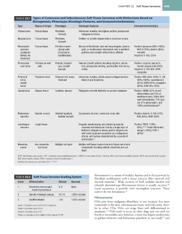
TABLE 22.1 Types of Cutaneous and Subcutaneous Soft Tissue Sarcomas with Distinctions Based on
Histogenesis, Phenotype, Histologic Features, and Immunohistochemistry
VetBooks.ir Type Tissue of Origin Phenotype Histologic Features Immunohistochemistry
Interwoven bundles, herringbone pattern, pronounced
Fibrous tissue
Fibroblast,
Fibrosarcoma
fibrocyte collagenous stroma
Myxosarcoma Fibrous tissue Fibroblast, Stellate- or spindle-shaped cells in mucinous stroma
fibrocyte
Pleomorphic Fibrous tissue Primitive mesen- Mixture of fibroblastic cells and karyomegalic, cytome- Positive: lysozyme (29%–100%),
sarcoma chymal cells galic, or multinucleate histiocytoid cells in storiform MHC II (70%), desmin (86%),
(malignant (fibroblast or patterns with variable inflammatory infiltrate vimentin
fibrous his- myofibroblast) Negative: S-100, CD18
tiocytoma)
Perivascular Perivascular wall Pericyte, myoperi- Vascular growth patterns including staghorn, placen- Positive: calponin, pan actin,
wall tumor cells cyte, smooth toid, perivascular whirling, and bundles from tunica smooth muscle actin (50%)
myocyte media Negative: S-100, NSE, GFAP,
myoglobin
Peripheral Peripheral nerve Schwann cell, neuro- Interwoven bundles, whorls around collagen bundles, Positive: NSE (45%–82%), S-100
nerve fibroblast Antoni A and B patterns (50%–100%), neurofilament
sheath (82%), NGFR (47%), myoglobin
tumor (64%), GFAP (0%–35%)
Liposarcoma Adipose tissue Lipoblast, lipocyte Polygonal cells with distinctly vacuolated cytoplasm Positive: MDM2 (67% of well-
differentiated and 75% of
dedifferentiated), CDK4 (88%
well-differentiated, 71% myx-
oid, 67% pleomorphic, and
100% dedifferentiated) 84
Rhabdomyo- Skeletal muscle Skeletal myoblast, Cytoplasmic striation, racket and strap cells Positive: desmin, S-100 (75%),
sarcoma skeletal mycoyte NSE (50%), GFAP (50%)
Lymphangio- Lymph tissue Irregular, anastomosing, and arborizing vascular Positive: PROX-1 (80%–
sarcoma channels and trabeculae lined by a single layer of 88%), 90,91 Factor VIII-related
flattened, elongate to plump spindle-shaped cells antigen (100%), LYVE-1
with scant cytoplasm supported on a collagenous (80%) 91
stroma; with lumina characterized by a paucity of
erythrocytes. 91
Mesenchy- Any mesenchy- Multiple cell types Multiple soft tissue mesenchymal cell types and matrix
moma mal tissue components including osteoid, chondroid, and col-
lagen
GFAP, Glial fibrillary acidic protein; LYVE-1, lymphatic vessel endothelial receptor-1; MDM2, mouse double minute 2 homolog; MHC, major histocompatibility complex; NGFR, nerve growth factor receptor;
NSE, neuron specific enolase; PROX-1, prospero-related homeobox gene 1.
Modified from Dennis et al, Vet Pathol, 2011. 12,84,90,91
fibromatosis is a variant of nodular fasciitis and is characterized by
TABLE 22.2 Soft Tissue Sarcoma Grading System
fibroblast proliferation with a dense reticular fiber network and
27
Score Differentiation Mitosis a Necrosis mucoid material. Wide excision of both nodular fasciitis and
28
infantile desmoid-type fibromatosis lesions is usually curative.
1 Resembles normal adult 0–9 None Local recurrence is possible with incomplete resection. These
mesenchymal tissue
26
tumors do not metastasize.
2 Specific histologic subtype 10–19 <50% necrosis
Fibrosarcoma
3 Undifferentiated >20 >50% necrosis
FSAs arise from malignant fibroblasts in any location, but most
Grade I: Cumulative score of ≤4 for the 3 categories. commonly in the skin, subcutaneous tissue, and oral cavity. Simi-
Grade II: Cumulative score of 5–6. lar to other STSs, FSAs can range from well differentiated to
29
Grade III: Cumulative score of ≥7. anaplastic. FSAs tend to occur in older dogs and cats with no
a Mitosis is calculated as the number of mitotic figures/10 HPF. breed or sex predilection; however, a there was higher predilection
in golden retrievers and Doberman pinschers in one study and
30